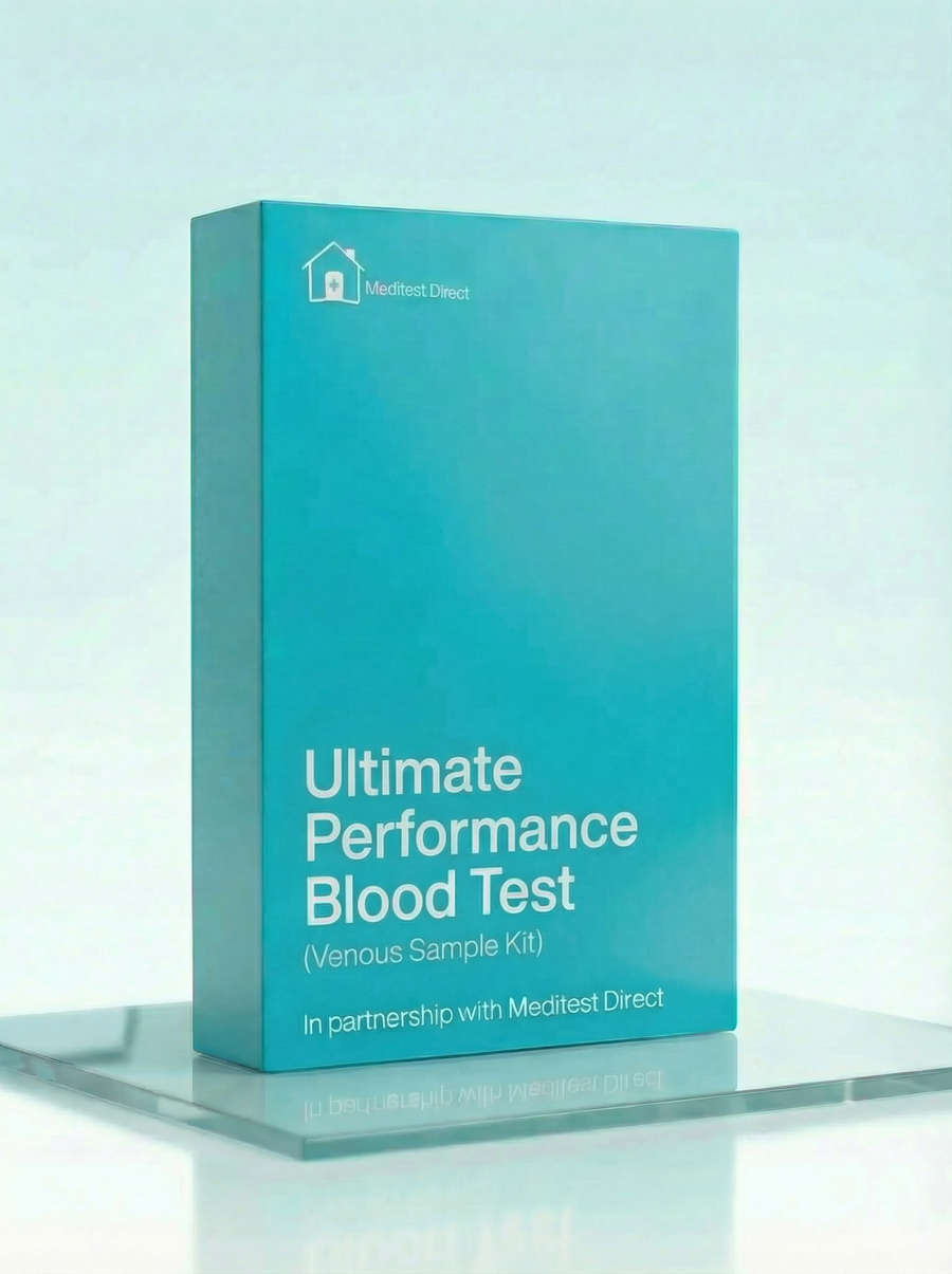
Ultimate Performance Blood Test Kit
£199 ✓ In Stock
Your sample goes to a UKAS accredited laboratory meeting ISO 15189 standards.
After you receive your order confirmation email, please reply with your date of birth.
How it works
Your testing journey
From order to results in four simple steps. Full transparency on where each step happens and what it costs.

Receive your kit by post
Dispatched same working day if ordered before 3pm. Royal Mail Tracked delivery, typically 1–3 working days. 90% of kits arrive within 24 hours.

Visit a partner clinic
Book a phlebotomy appointment at one of our 365+ UK partner clinics. Take your kit with you — the phlebotomist will collect your sample using the materials provided.
Phlebotomy fee applies (paid at clinic)
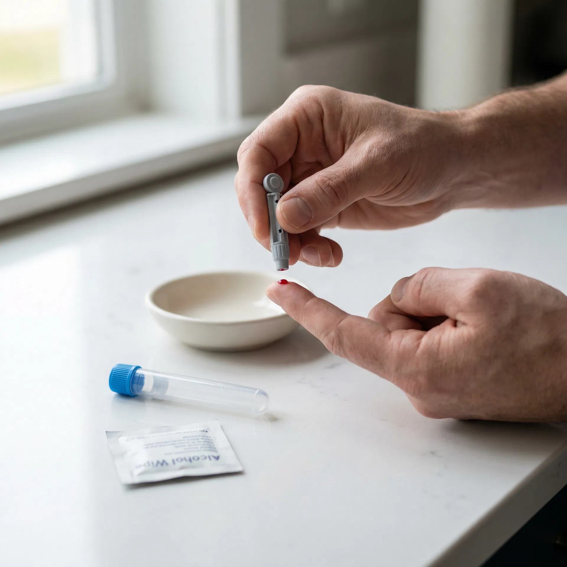
Venous blood draw at a clinic
A trained phlebotomist takes a small blood sample from a vein in your arm using the vacutainers provided in your kit. The appointment takes around 10 minutes.

Return by prepaid envelope
Seal your sample in the biohazard bag provided and drop it in any Royal Mail postbox using the prepaid Tracked 24 envelope. Post Monday–Thursday for best results.
Venous Blood Collection Kit
This kit is sent to you and taken to your chosen clinic. The phlebotomist will collect your sample using the materials provided.
- 1Vacutainer blood collection tubes
- 2Needle and butterfly needle
- 3Tourniquet
- 4Alcohol swab
- 5Cotton wool and gauze
- 6Adhesive plaster
- 7Biohazard specimen bag
- 8Prepaid return envelope (Royal Mail Tracked 24)
- 9Laboratory request form
- 10Instructions for the phlebotomist
Testosterone is the primary male sex hormone, crucial for muscle mass, bone density, red blood cell production, libido, and mood. Women also produce testosterone, just in smaller amounts. In men, normal range is roughly 8.6–29 nmol/L; levels below 8 nmol/L typically warrant investigation. Testosterone peaks in early morning and declines with age (about 1–2% per year after 30). Overtraining, poor sleep, excessive alcohol, and obesity can suppress testosterone. For accurate assessment, test between 7–10am. Two separate low readings are typically required before diagnosing hypogonadism. Results outside the normal range may need a follow-up with your GP.
SHBG is a protein that binds testosterone and oestradiol, controlling how much is "free" and biologically active. High SHBG means more testosterone is bound and unavailable—total testosterone might look normal while free testosterone is low. Low SHBG means more free testosterone. SHBG increases with age, hyperthyroidism, low body weight, and oestrogen; it decreases with obesity, hypothyroidism, insulin resistance, and androgen use. Understanding SHBG is essential for interpreting testosterone results accurately. Results outside the normal range may need a follow-up with your GP.
The Free Androgen Index estimates biologically active testosterone by calculating (Total Testosterone ÷ SHBG) × 100. It's useful when SHBG is abnormal—someone with high total testosterone but very high SHBG might have a normal or low FAI, explaining symptoms despite "normal" testosterone. In women, FAI above 5 suggests androgen excess (as seen in PCOS). In men, it helps identify those with low free testosterone despite borderline total testosterone. FAI is a calculated estimate—directly measured free testosterone is more accurate but more expensive. Results outside the normal range may need a follow-up with your GP.
Oestradiol is the primary oestrogen, important for bone health, cardiovascular protection, and reproductive function in both sexes. In women, levels vary dramatically through the menstrual cycle—testing on day 2–5 provides a baseline. In men, oestradiol is produced by converting testosterone via aromatase (mainly in fat tissue). Elevated oestradiol in men can cause gynaecomastia, water retention, and mood changes; very low oestradiol affects bone health and libido. Men on testosterone replacement therapy need oestradiol monitoring. Results outside the normal range may need a follow-up with your GP.
FSH is produced by the pituitary gland and stimulates egg development in women and sperm production in men. High FSH suggests the gonads aren't responding adequately—in women, this indicates menopause or premature ovarian insufficiency; in men, primary testicular failure. Low FSH can indicate pituitary problems. In women, FSH varies through the cycle—day 2–5 testing is standard for fertility assessment. FSH above 25 IU/L in women strongly suggests menopause. In men, FSH with testosterone helps distinguish primary from secondary hypogonadism. Results outside the normal range may need a follow-up with your GP.
LH triggers ovulation in women and stimulates testosterone production in men. Like FSH, it's produced by the pituitary. High LH with low testosterone in men indicates primary testicular failure; low LH with low testosterone suggests pituitary or hypothalamic problems. In women, the LH: FSH ratio helps diagnose PCOS (typically elevated LH). LH surges mid-cycle to trigger ovulation—timing matters for interpretation. Together with FSH and sex hormones, LH helps map the entire hypothalamic-pituitary-gonadal axis. Results outside the normal range may need a follow-up with your GP.
Prolactin is best known for stimulating breast milk production, but it also affects libido and reproductive function in both sexes. Elevated prolactin (hyperprolactinaemia) can suppress testosterone and cause erectile dysfunction in men, or menstrual irregularities in women. Causes include pituitary tumours (prolactinomas), certain medications (especially antipsychotics), hypothyroidism, and stress. Prolactin rises with stress, nipple stimulation, and sleep—morning testing after avoiding these is most accurate. Very high levels (above 5000 mU/L) strongly suggest prolactinoma. Results outside the normal range may need a follow-up with your GP.
Cortisol is the primary stress hormone, produced by the adrenal glands following a strong circadian rhythm—highest in early morning, lowest around midnight. It regulates metabolism, immune function, and stress response. Chronically elevated cortisol (Cushing's syndrome) causes weight gain, muscle weakness, and metabolic problems. Very low cortisol (Addison's disease) causes fatigue, low blood pressure, and salt cravings. For athletes, the testosterone:cortisol ratio can indicate overtraining—when cortisol rises relative to testosterone, recovery is impaired. Morning testing (7–9am) is standard. Results outside the normal range may need a follow-up with your GP.
DHEA-S (dehydroepiandrosterone sulphate) is produced by the adrenal glands and serves as a precursor to sex hormones. It's the most abundant steroid hormone in the body and declines steadily with age—levels at 70 are about 20% of levels at 20. Low DHEA-S can indicate adrenal insufficiency; very high levels in women may contribute to androgen excess symptoms. DHEA-S doesn't fluctuate much throughout the day, making it easier to interpret than cortisol. Some people supplement DHEA hoping to slow ageing, though evidence for benefits is limited. Results outside the normal range may need a follow-up with your GP.
IGF-1 is produced mainly by the liver in response to growth hormone and mediates most of growth hormone's effects on tissues. It's important for muscle growth, bone health, and tissue repair. Unlike growth hormone (which pulses throughout the day), IGF-1 levels are stable and reflect average growth hormone activity. Low IGF-1 may indicate growth hormone deficiency; very high levels can suggest acromegaly (growth hormone excess). For athletes, adequate IGF-1 supports recovery and adaptation. IGF-1 declines with age, caloric restriction, and protein deficiency. Results outside the normal range may need a follow-up with your GP.
TSH is produced by the pituitary gland and controls thyroid hormone production through a feedback loop. High TSH indicates the pituitary is working harder to stimulate an underactive thyroid (hypothyroidism); low TSH suggests an overactive thyroid (hyperthyroidism) or pituitary problems. TSH is the most sensitive screening test for thyroid dysfunction—it changes before Free T4 and Free T3 become abnormal. Normal range is typically 0.4–4.0 mU/L, though optimal may be narrower. TSH follows a circadian rhythm, peaking overnight and lowest in afternoon. Results outside the normal range may need a follow-up with your GP.
Free T4 measures the unbound, biologically active portion of thyroxine—the main hormone produced by the thyroid gland. T4 is a prohormone that converts to the more active T3 in tissues. Low Free T4 with high TSH confirms primary hypothyroidism; high Free T4 with low TSH confirms hyperthyroidism. Free T4 is more accurate than total T4 because it's not affected by binding protein levels (which vary with pregnancy, oestrogen, and liver disease). Normal range is typically 9–25 pmol/L. Results outside the normal range may need a follow-up with your GP.
Free T3 is the most metabolically active thyroid hormone, controlling metabolism, energy, heart rate, and body temperature. Most T3 is produced by converting T4 in peripheral tissues. Low Free T3 with normal T4 can occur in "sick euthyroid syndrome" during illness, fasting, or extreme stress—the body deliberately reduces metabolism. Some people with hypothyroid symptoms despite normal TSH and T4 may have poor T4-to-T3 conversion. Athletes in heavy training or caloric deficit may see lower Free T3. Results outside the normal range may need a follow-up with your GP.
Serum iron measures the amount of iron circulating in your blood bound to transferrin. It fluctuates significantly throughout the day and with recent food intake, making it less reliable than ferritin for assessing overall iron status. Morning levels are typically highest. Low serum iron can indicate iron deficiency but also occurs during inflammation or infection (the body sequesters iron). High serum iron may suggest iron overload, haemochromatosis, or recent iron supplementation. For athletes, iron is crucial for oxygen transport and energy production. Results outside the normal range may need a follow-up with your GP.
TIBC measures how much iron your blood could carry if transferrin were fully saturated. It reflects transferrin levels and responds inversely to iron stores—when iron is low, the body produces more transferrin (raising TIBC) to scavenge whatever iron is available. High TIBC with low serum iron and low ferritin is the classic pattern of iron deficiency. Low TIBC can indicate iron overload, chronic inflammation, or liver disease. TIBC doesn't fluctuate as much as serum iron, making it more reliable for trend monitoring. Results outside the normal range may need a follow-up with your GP.
Transferrin saturation is calculated by dividing serum iron by TIBC—it shows what percentage of your iron-carrying capacity is actually being used. Normal is typically 20–50%. Below 20% suggests iron deficiency; above 45% may indicate iron overload. Transferrin saturation above 45% in men or post-menopausal women warrants investigation for haemochromatosis, a genetic condition causing excessive iron absorption. Athletes, particularly endurance athletes and menstruating women, often have lower transferrin saturation and benefit from monitoring. Results outside the normal range may need a follow-up with your GP.
Ferritin is the body's iron storage protein and the best single marker of iron status. Low ferritin (below 30 µg/L) indicates depleted iron stores, often causing fatigue before anaemia develops. Many athletes aim for ferritin above 50 µg/L for optimal performance. However, ferritin is also an acute phase reactant—it rises with inflammation, infection, and liver disease, potentially masking underlying iron deficiency. Very high ferritin (above 300 in men, 200 in women) warrants investigation for haemochromatosis or other causes. Results outside the normal range may need a follow-up with your GP.
ALT is an enzyme found primarily in liver cells. When liver cells are damaged, ALT leaks into the bloodstream, making it a sensitive marker of liver injury. Elevated ALT can indicate fatty liver disease, hepatitis, medication toxicity, or alcohol-related damage. In athletes, intense exercise can temporarily raise ALT—particularly after endurance events or heavy resistance training—so timing your test matters. Chronically elevated ALT warrants investigation even if you feel fine, as liver disease is often silent in early stages. Results outside the normal range may need a follow-up with your GP.
AST is found in the liver, heart, muscles, and other tissues. Unlike ALT, elevated AST isn't specific to the liver—muscle damage from exercise can raise it significantly. Comparing AST to ALT helps determine the source: if both are elevated with AST higher than ALT, muscle damage or alcohol-related liver disease may be involved. Athletes often see elevated AST after hard training sessions, marathons, or contact sports. Resting for 48–72 hours before testing helps distinguish true liver issues from exercise-induced elevations. Results outside the normal range may need a follow-up with your GP.
GGT is an enzyme involved in the transport of amino acids and is particularly sensitive to alcohol consumption and bile duct problems. It's often the first liver enzyme to rise with excessive alcohol intake, making it useful for monitoring drinking habits. GGT can also be elevated by certain medications, obesity, and metabolic syndrome. Unlike ALT and AST, GGT isn't typically affected by exercise. A persistently elevated GGT in someone who doesn't drink may indicate fatty liver disease or warrant further investigation. Results outside the normal range may need a follow-up with your GP.
ALP is found in the liver, bones, kidneys, and digestive system. Elevated levels can indicate liver or bile duct problems, but also bone conditions—growing children and adolescents naturally have higher ALP due to bone development. In adults, elevated ALP alongside elevated GGT suggests a liver or biliary cause; elevated ALP with normal GGT points toward bone. Certain medications, pregnancy, and healing fractures can also raise ALP. It's a useful marker but often requires other tests to interpret properly. Results outside the normal range may need a follow-up with your GP.
Total protein measures the combined amount of albumin and globulins in your blood. These proteins perform essential functions: maintaining fluid balance, transporting substances, fighting infections, and clotting blood. Low total protein can indicate liver disease (reduced production), kidney disease (protein loss), malnutrition, or malabsorption. High levels may suggest chronic inflammation, infection, or certain blood cancers. Dehydration can falsely elevate total protein by concentrating the blood. For athletes, adequate protein intake supports recovery and performance. Results outside the normal range may need a follow-up with your GP.
Albumin is the most abundant protein in blood plasma, produced exclusively by the liver. It maintains oncotic pressure (keeping fluid in blood vessels), transports hormones, vitamins, and medications, and serves as a marker of nutritional status and liver function. Low albumin can indicate liver disease, kidney disease (nephrotic syndrome), malnutrition, or chronic illness. Because the liver has significant reserve capacity, albumin often doesn't drop until liver disease is advanced. Athletes with adequate nutrition typically have normal albumin levels. Results outside the normal range may need a follow-up with your GP.
Globulins are a diverse group of proteins including immunoglobulins (antibodies), transport proteins, and clotting factors. Elevated globulins often indicate chronic inflammation, infection, or immune system activation. Low globulins can suggest immune deficiency or protein loss. The globulin level is calculated by subtracting albumin from total protein. Chronic infections, autoimmune conditions, and certain cancers can elevate globulins. Athletes experiencing frequent infections might have low immunoglobulin levels worth investigating. Results outside the normal range may need a follow-up with your GP.
This ratio compares albumin to globulin levels and can help identify the cause of abnormal protein levels. A low ratio (below 1.0) may indicate overproduction of globulins (chronic inflammation, infection, autoimmune disease) or underproduction of albumin (liver disease, malnutrition). A high ratio is less common but can occur with underproduction of immunoglobulins. The ratio provides context that individual protein levels alone might miss, helping guide further investigation when abnormalities are detected. Results outside the normal range may need a follow-up with your GP.
Creatinine is a waste product from muscle metabolism, filtered out by the kidneys at a fairly constant rate. Elevated creatinine suggests the kidneys aren't filtering efficiently—but context matters. Muscular individuals naturally have higher creatinine due to greater muscle mass; this doesn't indicate kidney problems. Creatine supplementation can also raise creatinine levels. Acute dehydration temporarily elevates creatinine by concentrating the blood. Persistently elevated creatinine warrants investigation, but a single high reading in a well-hydrated, muscular athlete isn't necessarily concerning. Results outside the normal range may need a follow-up with your GP.
eGFR estimates how well your kidneys filter waste from blood, calculated from creatinine, age, sex, and ethnicity. It's expressed as mL/min/1.73m² and staged: above 90 is normal, 60–89 is mildly reduced, 30–59 is moderately reduced, and below 30 indicates severe impairment. The calculation can underestimate kidney function in very muscular individuals (who have high creatinine from muscle, not kidney problems). If your eGFR seems low but you're fit and healthy, a cystatin C-based eGFR may give a more accurate picture. Results outside the normal range may need a follow-up with your GP.
Urea is a waste product from protein metabolism, produced in the liver and excreted by the kidneys. Elevated urea can indicate kidney impairment, dehydration, high protein intake, or gastrointestinal bleeding (blood is a protein source). Athletes on high-protein diets may have slightly elevated urea that's entirely normal for them. Very low urea might suggest low protein intake or liver problems. Unlike creatinine, urea levels fluctuate more with diet and hydration status, making it a less reliable solo marker of kidney function. Results outside the normal range may need a follow-up with your GP.
This ratio helps distinguish between different causes of elevated kidney markers. A high ratio (elevated urea with relatively normal creatinine) suggests pre-renal causes like dehydration, high protein intake, or gastrointestinal bleeding. A normal ratio with both elevated suggests intrinsic kidney disease. A low ratio might indicate liver disease (reduced urea production) or low protein intake. For athletes, understanding this ratio helps contextualise results that might otherwise seem concerning—dehydration after a race, for example, typically elevates the ratio. Results outside the normal range may need a follow-up with your GP.
Total cholesterol measures all the cholesterol in your blood—the sum of LDL, HDL, and other lipoproteins. It's a starting point for understanding cardiovascular risk, but the breakdown matters more than the total number alone. Your liver produces most of your cholesterol; dietary cholesterol has less impact than once thought. Levels can be influenced by genetics, diet, exercise, and medications. Athletes often have higher HDL which can elevate total cholesterol while actually reducing cardiovascular risk. Results outside the normal range may need a follow-up with your GP.
Low-density lipoprotein (LDL) carries cholesterol from your liver to cells throughout the body. When there's too much, it can deposit in artery walls, forming plaques that narrow blood vessels over time—this is why it's often called "bad" cholesterol. However, LDL particle size matters: small, dense LDL particles are more atherogenic than large, fluffy ones. Diet, genetics, and lifestyle all influence LDL levels. Statins and other medications can lower LDL when lifestyle changes aren't enough. Results outside the normal range may need a follow-up with your GP.
High-density lipoprotein (HDL) is often called "good" cholesterol because it transports cholesterol away from arteries back to the liver for disposal. Higher HDL levels are generally protective against cardiovascular disease. Regular aerobic exercise is one of the most effective ways to raise HDL—athletes typically have higher levels than sedentary individuals. Moderate alcohol consumption can also raise HDL, though this isn't a recommended strategy. Genetics play a significant role; some people naturally have very high or very low HDL regardless of lifestyle. Results outside the normal range may need a follow-up with your GP.
Non-HDL cholesterol is calculated by subtracting HDL from total cholesterol—it represents all the potentially atherogenic cholesterol in your blood, including LDL, VLDL, and other lipoproteins. Many cardiologists consider this a better predictor of cardiovascular risk than LDL alone because it captures a broader picture. Unlike LDL, non-HDL can be measured accurately without fasting, making it practical for routine screening. Lower is generally better, and the target depends on your overall cardiovascular risk profile. Results outside the normal range may need a follow-up with your GP.
This ratio divides your total cholesterol by your HDL cholesterol, providing a quick snapshot of cardiovascular risk. A lower ratio is better—ideally below 4, with below 3.5 considered optimal. The ratio captures the balance between "good" and "bad" cholesterol in a single number. Someone with moderately elevated total cholesterol but high HDL might have a better ratio (and lower risk) than someone with lower total cholesterol but very low HDL. It's a useful metric for tracking progress over time as you make lifestyle changes. Results outside the normal range may need a follow-up with your GP.
Triglycerides are the most common type of fat in your blood, stored in fat cells and released for energy between meals. Elevated triglycerides are associated with increased cardiovascular risk and often accompany metabolic syndrome, insulin resistance, and type 2 diabetes. Unlike cholesterol, triglycerides respond dramatically to recent food intake—that's why fasting is required for accurate measurement. Refined carbohydrates and alcohol can spike triglycerides more than dietary fat. Regular exercise and reducing sugar intake are the most effective ways to lower them. Results outside the normal range may need a follow-up with your GP.
HbA1c measures the percentage of haemoglobin that has glucose attached to it, reflecting your average blood sugar over the past 2–3 months. It's the gold standard for diabetes diagnosis and monitoring because it isn't affected by what you ate yesterday or whether you fasted. Normal is below 42 mmol/mol (6.0%); 42–47 indicates prediabetes; 48 or above suggests diabetes. For athletes, it's useful for understanding how training and diet affect long-term glucose control. Conditions affecting red blood cell turnover (like anaemia or recent blood loss) can skew results. Results outside the normal range may need a follow-up with your GP.
Vitamin D is essential for calcium absorption, bone health, immune function, and muscle performance. Most people in the UK are deficient, particularly in winter. Levels below 25 nmol/L indicate deficiency; 25–50 is insufficient; 50–75 is adequate; above 75 is optimal for most purposes. Athletes may benefit from levels above 100 nmol/L for performance and recovery. Vitamin D is produced in skin with sun exposure and found in oily fish, eggs, and fortified foods. Supplementation (typically 1000–4000 IU daily) is often necessary in northern latitudes. Results outside the normal range may need a follow-up with your GP.
Vitamin B12 is essential for nerve function, DNA synthesis, and red blood cell production. Deficiency causes fatigue, weakness, numbness, memory problems, and macrocytic anaemia. B12 comes almost exclusively from animal products—vegans and vegetarians must supplement. Absorption requires intrinsic factor from the stomach; deficiency can occur despite adequate intake in older adults, those with pernicious anaemia, or after gastric surgery. The body stores years' worth of B12, so deficiency develops slowly. Levels below 200 ng/L warrant investigation; symptoms can occur even with "normal" levels. Results outside the normal range may need a follow-up with your GP.
Folate (vitamin B9) is crucial for DNA synthesis, cell division, and red blood cell formation. Deficiency causes macrocytic anaemia similar to B12 deficiency. Folate is found in leafy greens, legumes, and fortified cereals. Unlike B12, the body doesn't store much folate—deficiency can develop within weeks of inadequate intake. Alcohol interferes with folate absorption. Folate supplementation is essential before and during pregnancy to prevent neural tube defects. Importantly, folate supplementation can mask B12 deficiency anaemia while neurological damage continues—both should be checked together. Results outside the normal range may need a follow-up with your GP.
Magnesium is involved in over 300 enzymatic reactions including energy production, muscle contraction, nerve function, and protein synthesis. Deficiency can cause muscle cramps, fatigue, weakness, and heart rhythm abnormalities. Serum magnesium only reflects about 1% of body stores—levels can appear normal despite tissue depletion. Athletes lose magnesium through sweat and may have higher requirements. Good sources include nuts, seeds, whole grains, and leafy greens. Magnesium absorption decreases with high calcium intake, alcohol, and certain medications. Results outside the normal range may need a follow-up with your GP.
Haemoglobin is the protein in red blood cells that carries oxygen from lungs to tissues and returns carbon dioxide. Low haemoglobin defines anaemia—causing fatigue, weakness, and reduced exercise capacity. Normal ranges differ by sex: roughly 130–170 g/L for men, 120–150 g/L for women. Endurance athletes may have slightly lower haemoglobin due to plasma volume expansion ("sports anaemia"), which isn't true anaemia. High haemoglobin can indicate dehydration, altitude acclimatisation, or blood disorders. For performance, haemoglobin is a key determinant of VO2max. Results outside the normal range may need a follow-up with your GP.
This measures the number of red blood cells per litre of blood. Red cells live about 120 days and are constantly produced by bone marrow. Low counts occur in anaemia from various causes—iron deficiency, B12 deficiency, chronic disease, or bone marrow problems. High counts can result from dehydration, living at altitude, chronic lung disease, or polycythaemia. Athletes training at altitude naturally develop higher red cell counts to improve oxygen delivery. Combined with haemoglobin and other indices, red cell count helps classify different types of anaemia. Results outside the normal range may need a follow-up with your GP.
White blood cells are your immune system's soldiers. Elevated counts typically indicate infection, inflammation, or stress—the body mobilises white cells to fight threats. Very high counts can suggest leukaemia or other blood cancers. Low counts may indicate viral infection, autoimmune disease, bone marrow problems, or certain medications. Intense exercise causes transient elevation that returns to baseline within hours. Chronic overtraining can suppress white cell counts, potentially explaining increased infection susceptibility in overtrained athletes. Results outside the normal range may need a follow-up with your GP.
Platelets are cell fragments essential for blood clotting—they aggregate at wound sites to form the initial plug that stops bleeding. Low platelet counts (thrombocytopenia) increase bleeding risk and can result from viral infections, autoimmune conditions, medications, or bone marrow problems. High counts (thrombocytosis) may be reactive (due to inflammation, infection, or iron deficiency) or indicate a bone marrow disorder. Exercise doesn't significantly affect platelet counts. Normal range is typically 150–400 × 10⁹/L. Results outside the normal range may need a follow-up with your GP.
MCV measures the average size of your red blood cells in femtolitres (fL). It's crucial for classifying anaemia: low MCV (microcytic) suggests iron deficiency or thalassaemia; high MCV (macrocytic) suggests B12 or folate deficiency, alcohol excess, or thyroid problems; normal MCV (normocytic) with anaemia suggests chronic disease, acute blood loss, or bone marrow issues. MCV doesn't change quickly—it reflects the average over red cells' 120-day lifespan. Normal range is typically 80–100 fL. Results outside the normal range may need a follow-up with your GP.
MCH measures the average amount of haemoglobin in each red blood cell, expressed in picograms. It generally tracks with MCV—small cells (low MCV) contain less haemoglobin (low MCH), and large cells contain more. Low MCH (hypochromic) is characteristic of iron deficiency anaemia; high MCH occurs with B12 or folate deficiency. MCH is calculated by dividing total haemoglobin by red cell count. Combined with MCV and MCHC, it helps characterise the type of anaemia present. Results outside the normal range may need a follow-up with your GP.
MCHC measures the average concentration of haemoglobin in red blood cells—essentially how "packed" with haemoglobin each cell is. Low MCHC indicates hypochromic cells (pale, under-filled with haemoglobin), classic in iron deficiency. High MCHC is relatively rare and can indicate spherocytosis (a condition where red cells are abnormally shaped) or laboratory error. MCHC is more stable than MCV and MCH because it's a ratio rather than absolute value. It's particularly useful for detecting early iron deficiency. Results outside the normal range may need a follow-up with your GP.
RDW measures the variation in red blood cell size—how different your cells are from each other. Elevated RDW (anisocytosis) suggests the bone marrow is producing cells of inconsistent sizes, which occurs during recovery from anaemia, in mixed nutritional deficiencies, or with certain blood disorders. Iron deficiency anaemia typically shows high RDW (small, variable cells), while thalassaemia trait shows normal RDW (small but uniform cells). High RDW has also been associated with increased cardiovascular and mortality risk. Results outside the normal range may need a follow-up with your GP.
Neutrophils are the most abundant white blood cells and the first responders to bacterial infections. They engulf and destroy pathogens. Elevated neutrophils (neutrophilia) typically indicate bacterial infection, inflammation, stress, or corticosteroid use. Low neutrophils (neutropenia) increase infection risk and can result from viral infections, autoimmune conditions, certain medications, or bone marrow problems. Exercise causes transient neutrophilia that resolves within hours. Chronic overtraining may suppress neutrophil function. Results outside the normal range may need a follow-up with your GP.
Lymphocytes include T cells, B cells, and natural killer cells—the adaptive immune system's workhorses. Elevated lymphocytes (lymphocytosis) often indicate viral infection (glandular fever, flu, COVID-19) or, less commonly, lymphoma or leukaemia. Low lymphocytes (lymphopenia) can result from HIV, corticosteroid use, severe stress, or autoimmune conditions. Intense exercise temporarily suppresses lymphocyte counts, potentially creating an "open window" for infection—one reason athletes get sick after hard races. Results outside the normal range may need a follow-up with your GP.
Haematocrit measures the percentage of blood volume occupied by red blood cells. Normal is roughly 40–54% for men, 36–48% for women. Low haematocrit indicates anaemia or overhydration; high haematocrit suggests dehydration, polycythaemia, or altitude adaptation. Endurance athletes often have lower haematocrit due to expanded plasma volume—this isn't anaemia but a beneficial adaptation improving blood flow. Very high haematocrit (above 50%) increases blood viscosity and cardiovascular risk. In cycling, haematocrit above 50% triggers regulatory scrutiny. Results outside the normal range may need a follow-up with your GP.
CRP is an acute phase protein produced by the liver in response to inflammation. Levels can rise dramatically (10–100× normal) within hours of infection, injury, or surgery. It's useful for detecting and monitoring acute inflammation but doesn't indicate the source. CRP above 10 mg/L usually indicates significant inflammation—infection, autoimmune flare, or tissue damage. After intense exercise, CRP may rise temporarily. Very high CRP (above 100 mg/L) often indicates bacterial infection requiring urgent attention. Results outside the normal range may need a follow-up with your GP.
High-sensitivity CRP uses a more precise assay to detect low-grade chronic inflammation associated with cardiovascular risk. Levels below 1 mg/L indicate low risk; 1–3 mg/L is moderate; above 3 mg/L is higher risk. Unlike standard CRP, hs-CRP is useful when you're not acutely ill—it reflects smouldering background inflammation linked to atherosclerosis. Lifestyle factors like obesity, smoking, and poor diet elevate hs-CRP; exercise and weight loss lower it. If hs-CRP is above 10, acute illness is likely and the test should be repeated later. Results outside the normal range may need a follow-up with your GP.
Platelets are cell fragments essential for blood clotting—they aggregate at wound sites to form the initial plug that stops bleeding. Low platelet counts (thrombocytopenia) increase bleeding risk and can result from viral infections, autoimmune conditions, medications, or bone marrow problems. High counts (thrombocytosis) may be reactive (due to inflammation, infection, or iron deficiency) or indicate a bone marrow disorder. Exercise doesn't significantly affect platelet counts. Normal range is typically 150–400 × 10⁹/L. Results outside the normal range may need a follow-up with your GP.
MPV measures the average size of your platelets. Larger platelets are generally younger and more reactive—they contain more granules and are more effective at clotting. High MPV with low platelet count suggests the bone marrow is working hard to replace destroyed platelets (as in immune thrombocytopenia). Low MPV with low platelets suggests bone marrow isn't producing enough. MPV can also be elevated in cardiovascular disease and diabetes. It's a useful marker for understanding platelet dynamics beyond simple count. Results outside the normal range may need a follow-up with your GP.
Uric acid is produced when the body breaks down purines (found in certain foods and produced naturally). Most uric acid dissolves in blood, passes through the kidneys, and leaves in urine. When levels are too high (hyperuricaemia), crystals can form in joints causing gout—intensely painful inflammatory arthritis, often affecting the big toe. High uric acid also increases kidney stone risk. Purine-rich foods (organ meats, shellfish, beer) and fructose can elevate levels. Some people have genetic tendencies toward high uric acid. Results outside the normal range may need a follow-up with your GP.
This test is for screening and information only — it is not a medical diagnosis or professional advice. Please have your results reviewed by a qualified doctor or healthcare provider who can explain what they mean for your personal health situation. If your results show anything outside the normal range, or if you're worried about your health, see your doctor as soon as you can. Don't change any medications or treatments based on these results alone — always talk to your healthcare provider first.
NO CLINICS, NO QUEUES, NO HASSLE
Four steps to clarity
 01
01
Pick your panel
Browse over 200 clinically designed test kits and choose the one that fits your goals.
 02
02
Kit to your door
Everything you need arrives in discreet packaging with step-by-step instructions inside.
 03
03
Collect your sample
Follow the simple instructions in your kit — whether it's a finger-prick at home or a venous draw at a partner clinic.
 04
04
Insights delivered
Clear, easy-to-understand results sent to you online with actionable health guidance.
Frequently asked questions
This test measures Total Cholesterol, LDL Cholesterol, HDL Cholesterol, Non-HDL Cholesterol, Total:HDL Ratio. Check the full biomarker list on this page for detailed descriptions.
Check the Special Instructions on this page. General rule: fast 8-12 hours if cholesterol/glucose/insulin included. Most hormone, vitamin, and antibody tests do not require fasting. Morning collection (7-10am) is preferred.
Follow the instructions in your kit. For finger-prick: warm hands, use lancet as directed, fill tube to marked line. For venous: attend a phlebotomy clinic with your lab form. Post same day, avoid Fridays/bank holidays.
Results are typically available within the timeframe shown on this page. You will receive a notification when ready to view online.
You may also be interested in

Sports Hormone Blood Test Kit
A 42-biomarker venous blood panel covering cholesterol status, clotting status, diabetes and other key health markers — ideal for athletes and active people tracking training adaptation, recovery, and performance.
Results estimated in 2 working days
42 biomarkers

Fitness Blood Test Kit
A 15-biomarker venous blood panel covering cholesterol status, hormones, inflammation and other key health markers — ideal for athletes and active people tracking training adaptation, recovery, and performance.
Results estimated in 4 working days
15 biomarkers

Advanced Fitness Blood Test Kit
A 29-biomarker venous blood panel covering adrenal hormones, cholesterol status, diabetes and other key health markers — ideal for athletes and active people tracking training adaptation, recovery, and performance.
Results estimated in 4 working days
29 biomarkers

Driver's Tiredness Blood Test
A 6-biomarker venous blood panel covering inflammation, iron status, thyroid hormones and vitamins — ideal for investigating ongoing tiredness, low energy, or suspected medical causes.
Results estimated in 5 working days
6 biomarkers