Tiredness and Fatigue Blood Test
£78 ✓ In Stock
Your sample goes to a UKAS accredited laboratory meeting ISO 15189 standards.
After you receive your order confirmation email, please reply with your date of birth.
How it works
Your testing journey
From order to results in four simple steps. Full transparency on where each step happens and what it costs.

Receive your kit by post
Dispatched same working day if ordered before 3pm. Royal Mail Tracked delivery, typically 1–3 working days. 90% of kits arrive within 24 hours.

Visit a partner clinic
Book a phlebotomy appointment at one of our 365+ UK partner clinics. Take your kit with you — the phlebotomist will collect your sample using the materials provided.
Phlebotomy fee applies (paid at clinic)
Venous blood draw at a clinic
A trained phlebotomist takes a small blood sample from a vein in your arm using the vacutainers provided in your kit. The appointment takes around 10 minutes.

Return by prepaid envelope
Seal your sample in the biohazard bag provided and drop it in any Royal Mail postbox using the prepaid Tracked 24 envelope. Post Monday–Thursday for best results.
Venous Blood Collection Kit
This kit is sent to you and taken to your chosen clinic. The phlebotomist will collect your sample using the materials provided.
- 1Vacutainer blood collection tubes
- 2Needle and butterfly needle
- 3Tourniquet
- 4Alcohol swab
- 5Cotton wool and gauze
- 6Adhesive plaster
- 7Biohazard specimen bag
- 8Prepaid return envelope (Royal Mail Tracked 24)
- 9Laboratory request form
- 10Instructions for the phlebotomist
Thyroid stimulating hormone (TSH) is produced by the pituitary gland to regulate the thyroid. TSH levels reflect the body's assessment of thyroid hormone status: when thyroid hormones are low, TSH rises to stimulate more production; when thyroid hormones are high, TSH falls to slow production. This feedback loop makes TSH the most sensitive initial marker for thyroid dysfunction. An elevated TSH indicates that the thyroid is underperforming (hypothyroidism)—the pituitary is working harder to stimulate a sluggish thyroid. Hypothyroidism is a very common cause of fatigue, affecting around 2% of the UK population (more common in women and with increasing age). Other symptoms include weight gain, feeling cold, constipation, dry skin, hair loss, and depression. A low TSH indicates that the thyroid is overactive (hyperthyroidism)—the pituitary is backing off because thyroid hormones are already high. Hyperthyroidism can also cause fatigue, along with weight loss, anxiety, tremor, palpitations, and heat intolerance. Normal TSH is typically 0.4-4.0 mU/L, though "optimal" may be narrower (0.5-2.5 mU/L). Mildly elevated TSH (4-10 mU/L) is termed "subclinical hypothyroidism" and may or may not require treatment depending on symptoms and other factors. TSH should be interpreted alongside Free T4 for a complete picture. Results outside the normal range may need a follow-up with your GP.
Free T4 (free thyroxine) measures the unbound, biologically active portion of the thyroid hormone thyroxine. Most T4 in blood is bound to proteins and inactive; only the "free" fraction (about 0.03%) is available to enter cells and exert effects. T4 is the main hormone produced by the thyroid gland, though it must be converted to the more active T3 (triiodothyronine) within cells to have its full effect. T4 is the body's main metabolic regulator—it affects virtually every tissue and increases overall metabolic rate, heat production, heart rate, protein synthesis, and energy levels. Low Free T4 (with high TSH) confirms hypothyroidism and explains fatigue through reduced cellular metabolism. High Free T4 (with low TSH) confirms hyperthyroidism. In central hypothyroidism (rare), both TSH and Free T4 are low due to pituitary or hypothalamic problems. Normal Free T4 is typically 9-25 pmol/L. Interpreting Free T4 requires considering TSH alongside it: a low-normal Free T4 with a high TSH suggests early or subclinical hypothyroidism. Free T4 in the "normal" range doesn't necessarily mean thyroid function is optimal—some individuals feel better with Free T4 in the upper half of the reference range. Results outside the normal range may need a follow-up with your GP.
Serum iron measures the amount of iron circulating in your blood, bound to the transport protein transferrin. Iron is essential for making haemoglobin (the oxygen-carrying protein in red blood cells) and myoglobin (the equivalent protein in muscles), as well as for numerous enzymes involved in energy production, DNA synthesis, and other vital functions. Without adequate iron, cells cannot produce energy efficiently, leading to fatigue. Serum iron on its own is difficult to interpret because it fluctuates significantly throughout the day, in response to recent meals (particularly iron-rich foods), and due to many other factors. A single low serum iron reading doesn't necessarily mean iron deficiency, and a normal reading doesn't rule it out. This is why serum iron is always interpreted alongside other iron markers (TIBC, transferrin saturation, and ferritin) to get a complete picture of iron status. Normal serum iron is typically 10-30 µmol/L, though ranges vary between laboratories. Low serum iron with high TIBC, low transferrin saturation, and low ferritin indicates iron deficiency. Low serum iron with normal or low TIBC may indicate anaemia of chronic disease (where iron is present in the body but sequestered and unavailable). High serum iron may indicate iron overload conditions like haemochromatosis. Results outside the normal range may need a follow-up with your GP.
Total iron-binding capacity (TIBC) measures the maximum amount of iron that could be bound by proteins in your blood—primarily transferrin, the main iron transport protein. TIBC is an indirect measure of transferrin levels: the more transferrin available, the higher the iron-binding capacity. Your body regulates transferrin production based on iron status. When iron stores are low, the liver produces more transferrin to maximise iron capture and transport from the diet—so TIBC increases. When iron stores are adequate or high, less transferrin is needed—so TIBC decreases. This makes TIBC a useful indicator of iron stores when interpreted alongside serum iron and ferritin. Normal TIBC is typically 45-80 µmol/L. High TIBC (with low serum iron and low ferritin) strongly suggests iron deficiency—the body is ramping up its iron transport capacity because stores are depleted. Low TIBC may be seen in iron overload (haemochromatosis), chronic inflammation, liver disease, or malnutrition. Interpreting TIBC alongside other iron markers gives a much clearer picture than any single test. Results outside the normal range may need a follow-up with your GP.
Transferrin saturation is a calculated percentage showing what proportion of the body's iron-carrying capacity is currently being used. It's calculated by dividing serum iron by TIBC and multiplying by 100. This ratio provides a snapshot of how much iron is actually being transported relative to the maximum capacity available. Normal transferrin saturation is typically 20-50%. Low transferrin saturation (below 20%) indicates that less iron is being transported than the body's capacity allows—this is typical of iron deficiency. Very low transferrin saturation (<15-16%) strongly suggests iron deficiency and correlates with impaired iron delivery to tissues, even before anaemia develops. High transferrin saturation (above 45-50%) suggests iron overload—more iron is being transported than is healthy. Very high saturation (>60%) is concerning for haemochromatosis and warrants further investigation. Transferrin saturation is particularly useful because it can detect both iron deficiency and iron overload. It's less affected by inflammation than ferritin alone, making it valuable in situations where ferritin might be misleadingly elevated. Results outside the normal range may need a follow-up with your GP.
Ferritin is the body's primary iron storage protein. It holds iron in a soluble, non-toxic form in cells throughout the body (particularly the liver, spleen, and bone marrow) and releases it when needed. Measuring ferritin in blood gives an indirect but generally reliable indication of total body iron stores—a low ferritin almost always means low iron stores, making it the most specific marker for iron deficiency. Low ferritin is one of the most common and easily treatable causes of fatigue. Even ferritin levels that are technically "within range" but at the lower end (e.g., 15-30 µg/L) may be associated with fatigue symptoms, particularly in menstruating women. Many practitioners now aim for ferritin levels of at least 50 µg/L in patients with fatigue symptoms, as this is associated with better energy and exercise tolerance, particularly in athletes and those with heavy periods. Normal ferritin ranges are typically 15-300 µg/L for men and 15-200 µg/L for women, but optimal levels for wellbeing may be higher. However, ferritin is an acute phase reactant—it rises during inflammation, infection, and liver disease—so a "normal" ferritin in the context of illness doesn't rule out underlying iron deficiency. Very high ferritin (>500-1000 µg/L) requires investigation for iron overload, liver disease, inflammation, or haematological conditions. Results outside the normal range may need a follow-up with your GP.
Vitamin D is not truly a vitamin but a steroid hormone that the body produces when skin is exposed to UVB sunlight. The liver converts this to 25-hydroxyvitamin D (25-OH vitamin D), which is the main circulating form and the best indicator of overall vitamin D status. This is the form measured in blood tests. Vitamin D plays crucial roles beyond bone health: it affects muscle function, immune regulation, mood, and energy metabolism. Vitamin D deficiency is extremely common in the UK due to limited sunlight exposure, particularly from October to March when UVB radiation is insufficient for vitamin D synthesis regardless of time spent outdoors. People with darker skin, those who cover their skin for cultural or religious reasons, and people who spend little time outdoors are at particular risk. Symptoms of deficiency include fatigue, muscle weakness, muscle aches, bone pain, frequent infections, and low mood. Many people have no obvious symptoms despite significant deficiency. Vitamin D status is classified as: deficient (<25 nmol/L), insufficient (25-50 nmol/L), adequate (50-75 nmol/L), and optimal (>75 nmol/L). For those with fatigue, aiming for levels above 75 nmol/L may be beneficial. UK guidelines recommend that everyone consider taking a vitamin D supplement (10 µg/400 IU daily) during autumn and winter; those at risk of deficiency should supplement year-round. Higher doses may be needed to correct deficiency. Results outside the normal range may need a follow-up with your GP.
C-reactive protein (CRP) is an acute phase protein produced by the liver in response to inflammation anywhere in the body. High sensitivity CRP (hs-CRP) is measured using a more sensitive assay that can detect lower levels of inflammation than standard CRP tests. This makes it useful for detecting chronic low-grade inflammation that might not show up on a standard CRP test. Chronic inflammation is increasingly recognised as a contributor to fatigue. Inflammatory cytokines (the signalling molecules that trigger CRP production) directly affect the brain, causing symptoms that collectively resemble fatigue: low energy, reduced motivation, poor concentration, disturbed sleep, and mood changes. This "sickness behaviour" is an evolved response to infection but can become problematic when inflammation persists without an acute infection. Conditions associated with chronic low-grade inflammation include obesity, poor metabolic health, autoimmune conditions, chronic infections, and some mood disorders. Normal hs-CRP is generally below 3 mg/L, with lower levels (<1 mg/L) being optimal. Mild elevations (3-10 mg/L) may indicate chronic low-grade inflammation; very high levels (>10 mg/L) usually indicate acute infection or significant inflammation and the test should be repeated when you're fully recovered. If hs-CRP is elevated without obvious cause, it's worth investigating potential sources of inflammation and considering whether lifestyle factors (diet, weight, sleep, stress) might be contributing. Results outside the normal range may need a follow-up with your GP.
This test is for screening and information only — it is not a medical diagnosis or professional advice. Please have your results reviewed by a qualified doctor or healthcare provider who can explain what they mean for your personal health situation. If your results show anything outside the normal range, or if you're worried about your health, see your doctor as soon as you can. Don't change any medications or treatments based on these results alone — always talk to your healthcare provider first.
NO CLINICS, NO QUEUES, NO HASSLE
Four steps to clarity
 01
01
Pick your panel
Browse over 200 clinically designed test kits and choose the one that fits your goals.
 02
02
Kit to your door
Everything you need arrives in discreet packaging with step-by-step instructions inside.
 03
03
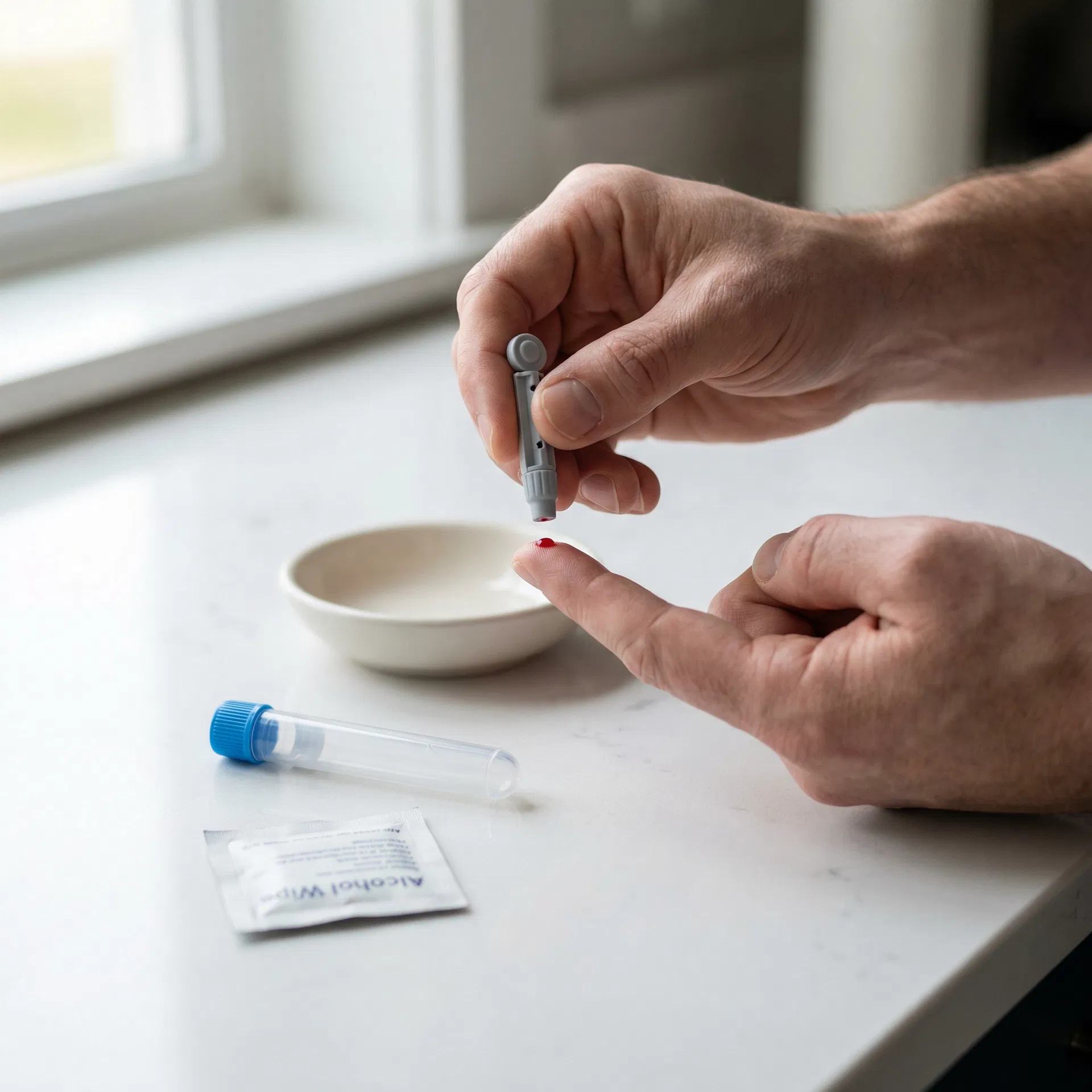
Collect your sample
Follow the simple instructions in your kit — whether it's a finger-prick at home or a venous draw at a partner clinic.
 04
04
Insights delivered
Clear, easy-to-understand results sent to you online with actionable health guidance.
Frequently asked questions
Key biomarkers associated with fatigue: thyroid function (most common treatable cause), iron/ferritin, vitamin D, B12, and blood sugar markers. Identifies the most common medical causes of tiredness.
If glucose/insulin included, fast 8-12 hours. For thyroid/vitamin/hormone markers, no. Morning 7-10am preferred.
Normal results rule out the most common medical causes. If symptoms persist, discuss with your GP — further investigation (sleep studies, mental health assessment, dietary review) may be helpful.
You may also be interested in

Anaemia Profile Blood Test
A 21-biomarker venous blood panel covering red blood cells, white blood cells, clotting status and other key health markers — ideal for investigating fatigue, brain fog, tingling, or pernicious anaemia.
Results estimated in 2 working days
21 biomarkers

Advanced Tiredness and Fatigue Blood Test Kit
A 26-biomarker venous blood panel covering clotting status, diabetes, inflammation and other key health markers — ideal for investigating ongoing tiredness, low energy, or suspected medical causes.
Results estimated in 3 working days
26 biomarkers

Advanced Thyroid Function Blood Test Kit
A 10-biomarker venous blood panel covering thyroid hormones, autoimmunity, vitamins and other key health markers — ideal for comprehensive thyroid assessment including antibodies.
Results estimated in 2 working days
10 biomarkers

Ultimate Performance Blood Test Kit
A 56-biomarker venous blood panel covering cholesterol status, diabetes, liver function and other key health markers — ideal for athletes and active people tracking training adaptation, recovery, and performance.
Results estimated in 3 working days
56 biomarkers