
Essential Blood Test
£89 ✓ In Stock
Your sample goes to a UKAS accredited laboratory meeting ISO 15189 standards.
After you receive your order confirmation email, please reply with your date of birth.
How it works
Your testing journey
From order to results in four simple steps. Full transparency on where each step happens and what it costs.

Receive your kit by post
Dispatched same working day if ordered before 3pm. Royal Mail Tracked delivery, typically 1–3 working days. 90% of kits arrive within 24 hours.

Visit a partner clinic
Book a phlebotomy appointment at one of our 365+ UK partner clinics. Take your kit with you — the phlebotomist will collect your sample using the materials provided.
Phlebotomy fee applies (paid at clinic)
Venous blood draw at a clinic
A trained phlebotomist takes a small blood sample from a vein in your arm using the vacutainers provided in your kit. The appointment takes around 10 minutes.

Return by prepaid envelope
Seal your sample in the biohazard bag provided and drop it in any Royal Mail postbox using the prepaid Tracked 24 envelope. Post Monday–Thursday for best results.
Venous Blood Collection Kit
This kit is sent to you and taken to your chosen clinic. The phlebotomist will collect your sample using the materials provided.
- 1Vacutainer blood collection tubes
- 2Needle and butterfly needle
- 3Tourniquet
- 4Alcohol swab
- 5Cotton wool and gauze
- 6Adhesive plaster
- 7Biohazard specimen bag
- 8Prepaid return envelope (Royal Mail Tracked 24)
- 9Laboratory request form
- 10Instructions for the phlebotomist
Total protein measures the combined amount of albumin and globulins in your blood. These proteins perform essential functions including maintaining fluid balance (keeping water inside blood vessels through osmotic pressure), transporting hormones, vitamins, and medications through the bloodstream, fighting infections (immunoglobulins), clotting blood, and supporting tissue growth and repair. Low total protein can indicate malnutrition, malabsorption, liver disease (reduced production), kidney disease (protein loss in urine), or chronic illness. High total protein may indicate dehydration (concentrating the proteins), chronic inflammation, or rarely certain blood cancers like multiple myeloma that produce excess immunoglobulins. Total protein is a general screening marker—abnormal results prompt further investigation into which protein fraction is affected. Results outside the normal range may need a follow-up with your GP.
Albumin is the most abundant protein in blood plasma, accounting for about 50-60% of total blood protein. It's produced exclusively by the liver. Albumin has several crucial functions: it maintains oncotic pressure (keeping fluid inside blood vessels and preventing swelling), transports hormones, fatty acids, bilirubin, and medications, and acts as a buffer helping maintain blood pH. Low albumin (hypoalbuminaemia) can cause fluid to leak out of blood vessels into tissues, causing oedema (swelling). Causes include liver disease (reduced production), kidney disease—particularly nephrotic syndrome (albumin loss in urine), malnutrition, malabsorption, burns, and chronic inflammation. Albumin has a long half-life of about 20 days, so it reflects protein status over weeks rather than recent intake. In inflammation, albumin acts as a 'negative acute phase reactant'—levels fall as the liver prioritises producing inflammatory proteins. Results outside the normal range may need a follow-up with your GP.
Globulins are a diverse group of proteins in the blood, calculated by subtracting albumin from total protein. They include alpha globulins (transport proteins), beta globulins (including transferrin and complement), and gamma globulins (immunoglobulins/antibodies). Unlike albumin which is made only in the liver, globulins are produced by both the liver and the immune system. Elevated globulins often indicate immune system activation—the body producing more antibodies in response to infection, inflammation, or autoimmune disease. Significantly elevated levels can sometimes indicate multiple myeloma or other plasma cell disorders. Low globulins may suggest immunodeficiency, meaning reduced ability to fight infections. The albumin:globulin ratio is sometimes calculated; a low ratio (relatively high globulins) may indicate chronic infection, autoimmune disease, liver disease, or certain cancers. Results outside the normal range may need a follow-up with your GP.
Serum iron measures the amount of iron circulating in your blood, bound to the transport protein transferrin. Iron is an essential mineral needed to make haemoglobin (which carries oxygen in red blood cells), myoglobin (which stores oxygen in muscles), and numerous enzymes involved in energy production and DNA synthesis. Serum iron levels fluctuate significantly throughout the day and are influenced by recent dietary iron intake, making this a somewhat variable marker. For this reason, serum iron is rarely interpreted in isolation—it's most useful when considered alongside ferritin, TIBC, and transferrin saturation to give a complete picture of iron status. Low serum iron can indicate iron deficiency, chronic disease, or blood loss; high serum iron can indicate iron overload conditions like haemochromatosis, or recent iron supplementation. Results outside the normal range may need a follow-up with your GP.
Total iron-binding capacity measures the maximum amount of iron that can be bound by proteins in the blood, primarily transferrin. Transferrin is the main protein that transports iron through the bloodstream to tissues that need it. TIBC essentially tells us how much transferrin is available to bind iron. TIBC is useful for distinguishing between different causes of abnormal iron levels. In iron deficiency, the body produces more transferrin to try to capture and transport more iron, so TIBC is typically elevated. Conversely, in iron overload conditions (haemochromatosis), chronic disease, or inflammation, TIBC tends to be low. In anaemia of chronic disease—a common pattern seen with infections, inflammatory conditions, or malignancy—iron and TIBC are both low, which differs from true iron deficiency where TIBC would be high. Results outside the normal range may need a follow-up with your GP.
Transferrin saturation is calculated by dividing serum iron by TIBC and expressing it as a percentage. It represents how much of the available iron-carrying capacity is actually being used—essentially, how 'full' your transferrin is with iron. This is one of the most useful markers for identifying iron deficiency or iron overload. Normal transferrin saturation is typically 20-45%. Low saturation (below 20%) suggests iron deficiency—there isn't enough iron to fill the available transport capacity. High saturation (above 45-50%) can indicate iron overload and is an important early marker for hereditary haemochromatosis, a genetic condition causing excessive iron absorption that can damage the liver, heart, and pancreas if untreated. In haemochromatosis, transferrin saturation often rises before ferritin becomes significantly elevated. Results outside the normal range may need a follow-up with your GP.
Ferritin is a protein that stores iron inside cells throughout the body, particularly in the liver, spleen, and bone marrow. A small amount of ferritin circulates in the blood, and measuring this provides an excellent indication of total body iron stores. Ferritin is generally considered the single best marker for assessing whether someone is iron deficient. Low ferritin indicates depleted iron stores and is often the first marker to fall in developing iron deficiency, before haemoglobin drops and anaemia develops. Optimal ferritin for energy and wellbeing is generally considered to be 50-70 µg/L or above, though the laboratory 'normal' range starts lower. However, ferritin is also an acute phase reactant—it rises during inflammation, infection, and liver disease—so it can be misleadingly normal or even high despite underlying iron deficiency. Significantly elevated ferritin (above 300-500 µg/L) can indicate iron overload, inflammation, liver disease, or rarely certain cancers. Results outside the normal range may need a follow-up with your GP.
Bilirubin is a yellow-orange pigment produced when the liver breaks down old red blood cells, specifically the haem component of haemoglobin. The liver processes bilirubin, conjugating it to make it water-soluble, then excreting it in bile. Bile travels to the intestines where bilirubin gives faeces their characteristic brown colour. A small amount of bilirubin circulates in the blood. Elevated bilirubin causes jaundice—yellowing of the skin and eyes. This can occur when the liver is damaged (hepatitis, cirrhosis), when bile ducts are blocked (gallstones, tumours), or when red blood cells are being destroyed faster than normal (haemolytic anaemia). However, the most common cause of mildly elevated bilirubin is Gilbert's syndrome, a harmless genetic variation affecting 3-7% of the population that causes the liver to process bilirubin slightly more slowly. Gilbert's syndrome typically causes bilirubin levels of 20-50 µmol/L and requires no treatment. Results outside the normal range may need a follow-up with your GP.
Alkaline phosphatase (ALP) is an enzyme found throughout the body, with the highest concentrations in the liver, bile ducts, and bones. Smaller amounts are found in the intestines, kidneys, and placenta. ALP plays roles in bone mineralisation and bile duct function. Elevated ALP can indicate liver or bile duct problems (obstruction, primary biliary cholangitis, liver tumours) or bone disorders (Paget's disease, healing fractures, bone tumours, osteomalacia). To distinguish between liver and bone sources, ALP is interpreted alongside other liver enzymes—if ALT and GGT are also elevated, a liver source is likely; if they're normal, a bone source is more probable. ALP is naturally higher in children and adolescents (due to bone growth), during pregnancy, and can rise after fatty meals. Results outside the normal range may need a follow-up with your GP.
Alanine aminotransferase (ALT) is an enzyme found predominantly in liver cells, with smaller amounts in the heart, muscles, and kidneys. When liver cells are damaged or inflamed, ALT leaks into the bloodstream, making it a sensitive marker for liver inflammation and damage. Because ALT is found mainly in the liver, it's more specific to liver disease than some other enzymes. Common causes of elevated ALT include non-alcoholic fatty liver disease (NAFLD/MASLD)—now the most common cause in developed countries—alcohol-related liver disease, viral hepatitis, medication effects, and obesity. ALT can also rise temporarily after intense exercise. Persistently elevated ALT warrants investigation to identify the underlying cause. The most effective intervention for many people is addressing metabolic factors through weight loss, reducing alcohol, improving diet, and increasing physical activity. Results outside the normal range may need a follow-up with your GP.
Gamma-glutamyl transferase (GGT) is an enzyme found mainly in the liver, bile ducts, and kidneys. GGT plays a role in the transport of amino acids across cell membranes and in glutathione metabolism (an important antioxidant system). GGT is particularly sensitive to alcohol consumption and bile duct problems. GGT is elevated in approximately 75% of people who drink alcohol chronically, making it a useful marker for alcohol-related liver effects. It's also elevated in bile duct obstruction, fatty liver disease, and can be induced by certain medications (including some anticonvulsants). GGT helps distinguish between bone and liver causes of elevated ALP—if ALP is high but GGT is normal, the source is likely bone; if both are elevated, it's likely liver or biliary. Some individuals have naturally higher GGT due to genetic variation. Results outside the normal range may need a follow-up with your GP.
Urea is a waste product produced by the liver when it breaks down proteins and amino acids. Once produced, urea travels through the bloodstream to the kidneys, which filter it out and excrete it in urine. Measuring blood urea levels therefore reflects both liver function (production) and kidney function (excretion). Elevated urea most commonly indicates reduced kidney function—the kidneys aren't filtering waste products efficiently. However, urea can also rise with dehydration, high protein diets, gastrointestinal bleeding (blood is a protein source that gets digested), certain medications, and heart failure. Low urea may be seen with low protein diets, liver disease (reduced production), or overhydration. Urea is best interpreted alongside creatinine and eGFR for a complete picture of kidney function. Results outside the normal range may need a follow-up with your GP.
Creatinine is a chemical waste product produced from the normal breakdown of creatine phosphate in muscles. Creatinine production is relatively constant and proportional to muscle mass, making it a useful marker for kidney function. Like urea, creatinine is filtered out of the blood by the kidneys and excreted in urine. Elevated creatinine indicates reduced kidney function—the kidneys are unable to filter and excrete creatinine efficiently. However, creatinine levels are also affected by muscle mass, so naturally muscular individuals may have higher baseline levels, while those with low muscle mass (elderly, sedentary) may have lower levels that could mask early kidney disease. Intense exercise can temporarily raise creatinine. Creatinine is used to calculate eGFR, which adjusts for age and sex to give a more accurate picture of kidney function. Results outside the normal range may need a follow-up with your GP.
The estimated glomerular filtration rate assesses how well the kidneys are filtering waste from the blood. The glomeruli are tiny filtration units in the kidneys (about one million in each kidney) that filter blood to produce urine. eGFR estimates how much blood these filters process per minute, calculated using your creatinine level, age, and sex. eGFR is expressed in mL/min/1.73m² and is used to stage chronic kidney disease: Stage 1 (≥90) indicates normal or high function with other evidence of kidney damage; Stage 2 (60-89) indicates mildly reduced function; Stage 3a (45-59) mild-moderate reduction; Stage 3b (30-44) moderate-severe reduction; Stage 4 (15-29) severely reduced; Stage 5 (<15) kidney failure. Importantly, kidney function naturally declines with age, and a single borderline result doesn't necessarily indicate disease—patterns over time are more informative. Results outside the normal range may need a follow-up with your GP.
Total cholesterol measures all the cholesterol circulating in your blood. Cholesterol is an essential fat (lipid) that plays vital roles in the body—it's a key component of cell membranes, is used to manufacture hormones including testosterone, oestrogen, and cortisol, and is needed for vitamin D synthesis and bile acid production. The liver produces most of the cholesterol your body needs, with the remainder coming from dietary sources. While cholesterol is essential, the balance between different types matters for cardiovascular health. Total cholesterol on its own has limited value in assessing heart disease risk because it doesn't distinguish between protective HDL and potentially harmful LDL. You can have a high total cholesterol due to high HDL (protective) or high LDL (harmful)—two very different risk profiles. The detailed breakdown in this panel gives the complete picture. Results outside the normal range may need a follow-up with your GP.
LDL (low-density lipoprotein) cholesterol is often called 'bad cholesterol' because elevated levels drive atherosclerosis—the build-up of fatty plaques inside artery walls. LDL particles transport cholesterol from the liver to tissues throughout the body, but when levels are too high, excess LDL penetrates the arterial lining and becomes oxidised, triggering inflammation and plaque formation that narrows and stiffens arteries over time. Atherosclerosis can lead to heart attacks, strokes, and peripheral vascular disease. The process typically develops over decades, often without symptoms until a major event occurs. Reducing LDL through diet (reducing saturated fat, increasing fibre), exercise, weight management, and if necessary medication (statins) is one of the most effective ways to reduce cardiovascular risk. Current guidelines recommend LDL below 3.0 mmol/L for most adults, with lower targets for those at higher risk. Results outside the normal range may need a follow-up with your GP.
Non-HDL cholesterol is calculated by subtracting protective HDL cholesterol from your total cholesterol. This gives a single number representing all the potentially harmful cholesterol particles in your blood—not just LDL, but also VLDL (very low-density lipoprotein), IDL (intermediate-density lipoprotein), and other atherogenic remnant particles. These additional particles may be even more harmful than LDL in promoting atherosclerosis. Many cardiovascular experts now consider non-HDL cholesterol a superior risk marker compared to LDL alone because it captures the full atherogenic burden. It's also more reliable as it can be accurately measured without fasting. The recommended target for non-HDL cholesterol is below 4.0 mmol/L for most adults, with lower targets (below 2.5 mmol/L) for those at high cardiovascular risk. Results outside the normal range may need a follow-up with your GP.
HDL (high-density lipoprotein) cholesterol is called 'good cholesterol' because it performs reverse cholesterol transport—collecting excess cholesterol from tissues and artery walls and returning it to the liver for disposal or recycling. This process helps prevent cholesterol accumulation in arteries and protects against atherosclerosis. Higher HDL levels are associated with reduced cardiovascular risk, though the relationship is more complex than simply 'higher is better.' Levels above 1.0 mmol/L in men and 1.2 mmol/L in women are generally considered protective. Regular aerobic exercise is one of the most effective ways to raise HDL, along with maintaining a healthy weight, not smoking, and moderate alcohol consumption. Smoking significantly lowers HDL while also promoting LDL oxidation—a double cardiovascular harm. Results outside the normal range may need a follow-up with your GP.
The cholesterol: HDL ratio is calculated by dividing your total cholesterol by your HDL cholesterol. This ratio indicates what proportion of your total cholesterol is the protective HDL type versus potentially harmful types. It's a useful single-number summary of your lipid balance and is used in cardiovascular risk calculators such as QRISK to predict your 10-year risk of heart attack or stroke. A lower ratio indicates a more favourable lipid profile and reduced cardiovascular risk. An ideal ratio is below 4; a ratio between 4-6 indicates moderate risk; and a ratio above 6 indicates significantly increased cardiovascular risk. The ratio can be improved by either raising HDL (through exercise, weight loss, smoking cessation) or lowering total/LDL cholesterol (through diet, lifestyle, and if necessary medication). Results outside the normal range may need a follow-up with your GP.
Triglycerides are a type of fat that circulates in your blood, providing energy to cells. After eating, your body converts excess calories—whether from fat, carbohydrates, or protein—into triglycerides, which are then transported to fat cells for storage. Between meals, hormones release triglycerides from fat stores to provide energy. Elevated triglycerides are associated with increased cardiovascular risk and are often part of metabolic syndrome—a cluster of conditions including abdominal obesity, high blood pressure, elevated blood sugar, and abnormal cholesterol that significantly increases heart disease and diabetes risk. High triglycerides often reflect excess calorie intake, high sugar or refined carbohydrate consumption, excessive alcohol intake, or poorly controlled diabetes. The target is below 1.7 mmol/L. Triglycerides are most accurately measured after an 8-12 hour fast, as levels rise significantly after eating. Results outside the normal range may need a follow-up with your GP.
HbA1c (glycated haemoglobin) measures your average blood glucose control over the past 2-3 months. When glucose circulates in the blood, it attaches to haemoglobin in red blood cells through a process called glycation. The higher your average blood glucose, the more haemoglobin becomes glycated. Since red blood cells live for approximately 120 days, HbA1c provides a longer-term picture of glucose control than a single blood glucose measurement. HbA1c is the gold standard test for screening for and monitoring diabetes. Normal is below 42 mmol/mol (6.0%); prediabetes is 42-47 mmol/mol (6.0-6.4%); and diabetes is diagnosed at 48 mmol/mol (6.5%) or above. Prediabetes represents an important intervention window—lifestyle changes at this stage can reduce progression to diabetes by more than 50%. Uncontrolled diabetes damages blood vessels and nerves throughout the body, leading to complications including cardiovascular disease, kidney disease, eye damage, nerve damage, and poor wound healing. Results outside the normal range may need a follow-up with your GP.
Haemoglobin is the iron-containing protein inside red blood cells that carries oxygen from the lungs to every tissue in the body and returns carbon dioxide to the lungs for exhalation. It gives blood its red colour. Haemoglobin concentration is the primary measure used to diagnose anaemia and assess the blood's oxygen-carrying capacity. Low haemoglobin (anaemia) reduces oxygen delivery to tissues, causing fatigue, weakness, shortness of breath, dizziness, pale skin, and reduced exercise tolerance. The most common cause worldwide is iron deficiency, but anaemia can also result from vitamin B12 or folate deficiency, chronic disease, blood loss, bone marrow disorders, and inherited conditions like thalassaemia. High haemoglobin can occur with dehydration, chronic lung disease, living at high altitude, or polycythaemia (overproduction of red cells). Results outside the normal range may need a follow-up with your GP.
Haematocrit (HCT), also called packed cell volume (PCV), measures the percentage of blood volume occupied by red blood cells. If your haematocrit is 45%, that means 45% of your blood volume consists of red blood cells, with the remaining 55% being plasma and other components. Haematocrit closely parallels haemoglobin and is reduced in anaemia and elevated in polycythaemia or dehydration. Because it measures percentage rather than absolute amount, haematocrit is particularly affected by hydration status—dehydration concentrates blood cells (raising haematocrit), while overhydration dilutes them (lowering haematocrit). Very high haematocrit (above 50-55%) increases blood viscosity, making the blood 'thicker' and harder to pump, which can increase the risk of blood clots, stroke, and heart attack. Results outside the normal range may need a follow-up with your GP.
Red blood cell (RBC) count measures the number of red blood cells in a given volume of blood, typically expressed as cells per litre. Red blood cells are produced in the bone marrow and live for approximately 120 days before being recycled by the spleen. The hormone erythropoietin, produced mainly by the kidneys, regulates red blood cell production in response to oxygen levels. A low RBC count contributes to anaemia, though the size and haemoglobin content of the cells also matters. High RBC count (polycythaemia) can be a primary bone marrow disorder or secondary to chronic hypoxia (lung disease, sleep apnoea, high altitude), certain tumours that produce erythropoietin, or dehydration. Some athletes train at altitude or use altitude tents to naturally increase RBC count and improve endurance performance. Results outside the normal range may need a follow-up with your GP.
Mean cell volume measures the average size of your red blood cells, expressed in femtolitres (fL). Red blood cell size is important because different causes of anaemia produce characteristic cell sizes, helping to identify the underlying cause. A low MCV (microcytic anaemia, small cells) is typically caused by iron deficiency or thalassaemia trait. A high MCV (macrocytic anaemia, large cells) is commonly caused by vitamin B12 or folate deficiency, excessive alcohol consumption, certain medications, liver disease, or thyroid disorders. Normal MCV with anaemia (normocytic anaemia) suggests chronic disease, acute blood loss, kidney disease, or bone marrow problems. MCV is a key tool for narrowing down the cause of anaemia and directing further investigation. Results outside the normal range may need a follow-up with your GP.
Mean cell haemoglobin measures the average amount of haemoglobin contained within a single red blood cell, expressed in picograms (pg). This indicates how much oxygen-carrying haemoglobin each cell can hold. MCH generally correlates with MCV—smaller cells (low MCV) contain less haemoglobin (low MCH), and larger cells contain more. Low MCH (hypochromic cells, pale-looking on microscopy) is typical of iron deficiency anaemia where there isn't enough iron to make adequate haemoglobin. High MCH occurs in macrocytic anaemias where the larger cells contain more haemoglobin. MCH is used alongside MCV and MCHC to characterise anaemia and guide diagnosis. Results outside the normal range may need a follow-up with your GP.
Mean cell haemoglobin concentration measures the average concentration of haemoglobin within red blood cells, expressed as grams per litre (g/L). While MCH measures total haemoglobin per cell, MCHC indicates how densely packed that haemoglobin is within the cell volume. Low MCHC indicates hypochromic (pale) red cells, typically seen in iron deficiency anaemia and thalassaemia, where cells lack adequate haemoglobin relative to their size. High MCHC is less common and can occur in hereditary spherocytosis (where red cells are abnormally shaped and densely packed with haemoglobin) or as a laboratory artefact. MCHC tends to be more stable than MCH across different conditions. Results outside the normal range may need a follow-up with your GP.
Red cell distribution width measures the variation in size among your red blood cells. Normally, red blood cells are fairly uniform in size. RDW is expressed as a percentage—higher values indicate more variation (anisocytosis), meaning you have a mix of smaller and larger cells. Elevated RDW often indicates the bone marrow is producing cells of varying sizes, which can occur during recovery from anaemia (a mix of old small cells and new normal cells), combined deficiencies (e.g., iron plus B12), or bone marrow dysfunction. RDW can help distinguish between causes of microcytic anaemia: in iron deficiency, RDW is typically elevated (variable cell sizes), while in thalassaemia trait, RDW is often normal (uniformly small cells). Interestingly, elevated RDW has also been associated with increased cardiovascular risk and mortality, independent of anaemia. Results outside the normal range may need a follow-up with your GP.
White blood cells (WBCs), also called leukocytes, are the cells of your immune system that protect the body against infection and disease. Unlike red blood cells which are uniform, there are several types of white blood cells, each with specific functions. Total WBC count measures the combined number of all white cell types. Elevated WBC count (leukocytosis) most commonly indicates infection—the body is producing more white cells to fight invaders. Other causes include inflammation, stress, smoking, corticosteroid medication, and rarely leukaemia. Low WBC count (leukopenia) can occur with viral infections (which can temporarily suppress production), autoimmune conditions, bone marrow disorders, and certain medications including chemotherapy. The differential count (breakdown by cell type) helps identify which specific cells are affected. Results outside the normal range may need a follow-up with your GP.
Neutrophils are the most abundant type of white blood cell, typically comprising 40-70% of the total WBC count. They are the immune system's rapid response force—the first cells to arrive at sites of infection, where they engulf and destroy bacteria and fungi through a process called phagocytosis. Neutrophils form pus at infection sites. Elevated neutrophils (neutrophilia) typically indicate bacterial infection, but also occur with inflammation, stress, tissue damage (surgery, heart attack), smoking, and steroid use. Low neutrophils (neutropenia) significantly increases infection risk because these cells are critical for fighting bacterial infections. Causes include viral infections, certain medications, autoimmune conditions, and bone marrow disorders. Some people of African, Middle Eastern, or Caribbean descent have naturally lower neutrophil counts (benign ethnic neutropenia) without increased infection risk. Results outside the normal range may need a follow-up with your GP.
Lymphocytes are the second most common type of white blood cell and are central to the adaptive immune system—the targeted, specific immune response that 'remembers' previous infections. There are three main types: T cells (which coordinate immune responses and directly kill infected cells), B cells (which produce antibodies), and natural killer cells (which destroy virus-infected and cancerous cells). Elevated lymphocytes (lymphocytosis) are typically seen in viral infections—the opposite pattern to bacterial infections which raise neutrophils. Chronic lymphocytosis can occur with certain chronic infections and rarely lymphocytic leukaemia. Low lymphocytes (lymphopenia) can occur with acute infections, HIV, corticosteroid treatment, autoimmune conditions, and certain medications. Lymphocyte counts naturally vary and a single mild abnormality often resolves on repeat testing. Results outside the normal range may need a follow-up with your GP.
Monocytes are the largest type of white blood cell and typically comprise 2-8% of the WBC count. They circulate in the blood for a few days before migrating into tissues, where they mature into macrophages or dendritic cells. Macrophages engulf and digest pathogens, dead cells, and debris; dendritic cells present antigens to lymphocytes to activate specific immune responses. Elevated monocytes (monocytosis) can occur with chronic infections (tuberculosis, bacterial endocarditis), chronic inflammatory conditions, recovery from acute infections, and some blood cancers. The chronic low-grade inflammation that accompanies obesity and cardiovascular disease can also mildly elevate monocytes. Low monocytes are less common and can occur with bone marrow disorders or certain infections. Results outside the normal range may need a follow-up with your GP.
Eosinophils typically comprise 1-4% of white blood cells and are specialised for fighting parasitic infections and modulating allergic and inflammatory responses. They contain granules packed with toxic proteins that can kill parasites but can also damage host tissues when eosinophils are chronically activated, as in severe asthma or eosinophilic oesophagitis. Elevated eosinophils (eosinophilia) most commonly indicate allergic conditions (hay fever, eczema, asthma, drug allergies), parasitic infections, or autoimmune/inflammatory conditions. In developed countries where parasitic infections are uncommon, allergy is the most frequent cause. Marked or persistent eosinophilia warrants investigation. Low eosinophils are usually not significant and can occur during acute infections or with corticosteroid use. Results outside the normal range may need a follow-up with your GP.
Basophils are the rarest type of white blood cell, typically comprising less than 1% of the total WBC count. They contain granules filled with histamine, heparin, and other inflammatory mediators that are released during allergic reactions and parasitic infections. Basophils are closely related to mast cells in tissues. Because basophils are so rare, mild variations are usually not clinically significant. Elevated basophils (basophilia) can occur with allergic reactions, chronic inflammation, certain infections, hypothyroidism, and some myeloproliferative disorders (bone marrow conditions). Significantly elevated basophils alongside other abnormal counts may warrant haematological investigation. Low basophils are rarely significant. Results outside the normal range may need a follow-up with your GP.
C-reactive protein (CRP) is produced by the liver in response to inflammation anywhere in the body. The high-sensitivity CRP (hs-CRP) test can detect very low levels of inflammation that the standard CRP test would miss. This makes it useful for detecting low-grade chronic inflammation associated with cardiovascular disease, rather than the obvious inflammation of acute infections or injuries. Low-grade chronic inflammation damages blood vessel walls and contributes to atherosclerosis. Elevated hs-CRP is an independent risk factor for heart attack and stroke, even in people with normal cholesterol. An hs-CRP below 1 mg/L indicates low cardiovascular risk; 1-3 mg/L indicates moderate risk; and above 3 mg/L indicates higher risk. However, any acute illness, infection, or injury will temporarily raise CRP significantly, so this test should be taken when you're feeling well. If results are elevated, repeat testing after any acute cause has resolved. Results outside the normal range may need a follow-up with your GP.
Platelets (thrombocytes) are the smallest type of blood cell, produced in the bone marrow. They play an essential role in haemostasis—the process that stops bleeding when you're injured. When a blood vessel is damaged, platelets rapidly accumulate at the injury site, stick together, and form a plug that helps stop blood loss. They also release chemicals that activate the clotting cascade, leading to fibrin formation that reinforces the platelet plug. Low platelet counts (thrombocytopenia) can lead to easy bruising, prolonged bleeding from cuts, and in severe cases, spontaneous bleeding. Causes include bone marrow disorders, autoimmune conditions, viral infections, certain medications, and liver disease. High platelet counts (thrombocytosis) can increase the risk of abnormal clot formation and may be reactive (due to infection, inflammation, or iron deficiency) or indicate a bone marrow disorder. Results outside the normal range may need a follow-up with your GP.
Mean Platelet Volume measures the average size of your platelets. Platelet size provides information about platelet production in the bone marrow and platelet function. Younger, newly-released platelets tend to be larger and more active than older, smaller platelets. MPV is interpreted alongside platelet count. A high MPV with low platelet count suggests the bone marrow is producing larger platelets to compensate for increased platelet destruction or consumption—seen in conditions like immune thrombocytopenia (ITP). A low MPV with low platelet count may suggest reduced bone marrow production. Elevated MPV has also been associated with increased cardiovascular risk in some studies, as larger platelets may be more reactive and promote clot formation. Results outside the normal range may need a follow-up with your GP.
Uric acid is a waste product produced when the body breaks down purines—chemical compounds found naturally in the body and in certain foods including red meat, organ meats, shellfish, and alcohol (especially beer). Normally, uric acid dissolves in the blood, passes through the kidneys, and is excreted in urine. When uric acid levels become too high (hyperuricaemia), it can crystallise and deposit in joints, causing the intensely painful inflammatory condition known as gout. Gout typically affects the big toe first but can involve any joint. High uric acid can also form kidney stones. Beyond gout, elevated uric acid is increasingly recognised as a marker of metabolic syndrome and is associated with increased cardiovascular risk, kidney disease, and type 2 diabetes. Uric acid levels can be reduced through dietary modification (limiting purine-rich foods and alcohol), weight loss, staying well hydrated, and if necessary medication. Results outside the normal range may need a follow-up with your GP.
This test is for screening and information only — it is not a medical diagnosis or professional advice. Please have your results reviewed by a qualified doctor or healthcare provider who can explain what they mean for your personal health situation. If your results show anything outside the normal range, or if you're worried about your health, see your doctor as soon as you can. Don't change any medications or treatments based on these results alone — always talk to your healthcare provider first.
NO CLINICS, NO QUEUES, NO HASSLE
Four steps to clarity
 01
01
Pick your panel
Browse over 200 clinically designed test kits and choose the one that fits your goals.
 02
02
Kit to your door
Everything you need arrives in discreet packaging with step-by-step instructions inside.
 03
03
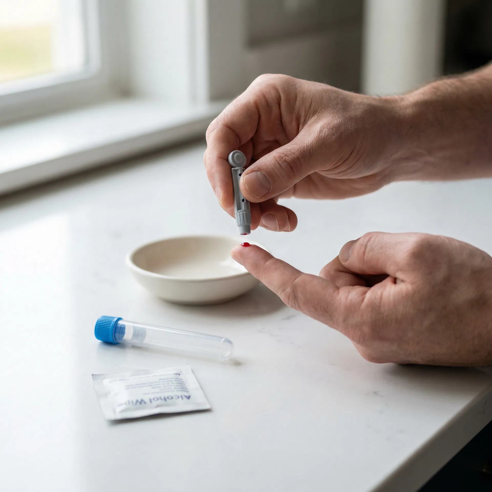
Collect your sample
Follow the simple instructions in your kit — whether it's a finger-prick at home or a venous draw at a partner clinic.
 04
04
Insights delivered
Clear, easy-to-understand results sent to you online with actionable health guidance.
Frequently asked questions
This test measures Total Cholesterol, LDL Cholesterol, Non-HDL Cholesterol, HDL Cholesterol, Total Cholesterol:HDL Ratio. Check the full biomarker list on this page for detailed descriptions of each marker and what it tells you about your health.
Check the Special Instructions section on this page. As a general rule, if the panel includes cholesterol, triglycerides, glucose, or insulin, fast for 8-12 hours. For most hormone, vitamin, and antibody tests, fasting is not required. Morning collection (7-10am) is preferred.
Follow the instructions in your kit. For finger-prick tests: warm your hands, use the lancet as directed, fill the tube to the marked line. For venous tests: attend a phlebotomy clinic with your laboratory request form. Post your sample the same day — avoid Fridays and bank holidays.
Results are typically available within the timeframe shown on this page. You will receive a notification when ready to view online. Results include reference ranges and guidance.
You may also be interested in

Core Health Blood Test Kit
A 7-biomarker venous blood panel covering cholesterol status and diabetes — ideal for investigating relevant symptoms or monitoring health.
Results estimated in 3 working days
7 biomarkers

Health and Lifestyle Blood Test Kit
A 19-biomarker venous blood panel covering cholesterol status, inflammation, iron status and other key health markers — ideal for investigating relevant symptoms or monitoring health.
Results estimated in 3 working days
19 biomarkers

Tiredness and Fatigue Blood Test
A 8-biomarker venous blood panel covering inflammation, iron status, thyroid hormones and vitamins — ideal for investigating ongoing tiredness, low energy, or suspected medical causes.
Results estimated in 3 working days
8 biomarkers

Vitamin D (25-OH) Blood Test Kit
A finger-prick blood test measuring your vitamin D (25-hydroxyvitamin D) level — the essential nutrient for bone health, immune function, and overall wellbeing, commonly deficient in the UK.
Results estimated in 2 working days
1 biomarkers