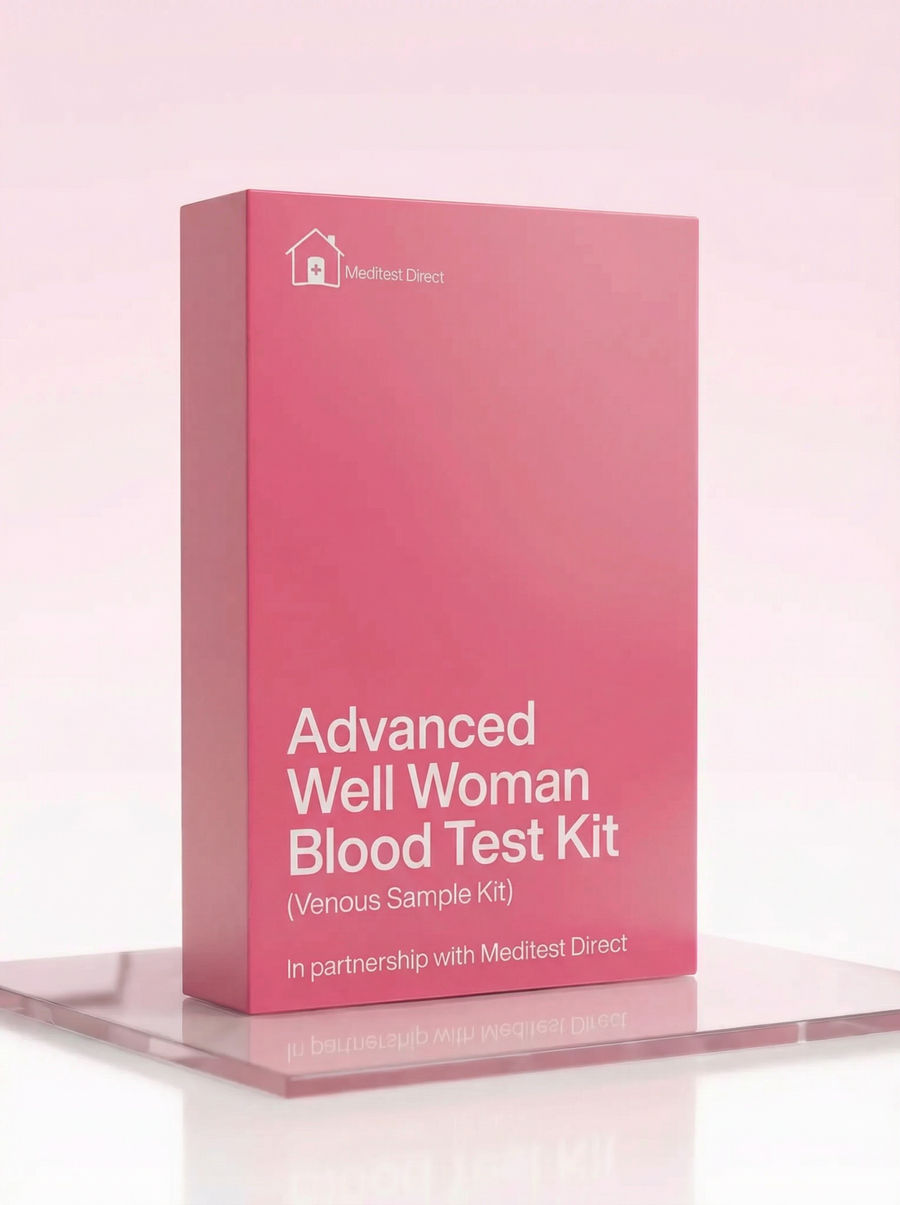
Advanced Well Woman Blood Test
£159 ✓ In Stock
Your sample goes to a UKAS accredited laboratory meeting ISO 15189 standards.
After you receive your order confirmation email, please reply with your date of birth.
How it works
Your testing journey
From order to results in four simple steps. Full transparency on where each step happens and what it costs.

Receive your kit by post
Dispatched same working day if ordered before 3pm. Royal Mail Tracked delivery, typically 1–3 working days. 90% of kits arrive within 24 hours.

Visit a partner clinic
Book a phlebotomy appointment at one of our 365+ UK partner clinics. Take your kit with you — the phlebotomist will collect your sample using the materials provided.
Phlebotomy fee applies (paid at clinic)
Venous blood draw at a clinic
A trained phlebotomist takes a small blood sample from a vein in your arm using the vacutainers provided in your kit. The appointment takes around 10 minutes.

Return by prepaid envelope
Seal your sample in the biohazard bag provided and drop it in any Royal Mail postbox using the prepaid Tracked 24 envelope. Post Monday–Thursday for best results.
Venous Blood Collection Kit
This kit is sent to you and taken to your chosen clinic. The phlebotomist will collect your sample using the materials provided.
- 1Vacutainer blood collection tubes
- 2Needle and butterfly needle
- 3Tourniquet
- 4Alcohol swab
- 5Cotton wool and gauze
- 6Adhesive plaster
- 7Biohazard specimen bag
- 8Prepaid return envelope (Royal Mail Tracked 24)
- 9Laboratory request form
- 10Instructions for the phlebotomist
FSH is produced by your pituitary gland and stimulates the ovaries to develop follicles containing eggs. In the first half of the menstrual cycle, FSH levels rise to promote follicle growth; one follicle becomes dominant and is released at ovulation. FSH levels vary throughout the cycle—they're lowest mid-cycle and highest just before and during menstruation. As women approach menopause and ovarian reserve declines, FSH levels rise as the pituitary works harder to stimulate the ovaries. Persistently elevated FSH (above 25-30 IU/L) with menopausal symptoms supports a diagnosis of menopause. For fertility assessment, FSH is best measured on day 2-5 of the cycle. Hormonal contraception suppresses FSH. Results outside the normal range may need a follow-up with your GP.
LH is produced by your pituitary gland and triggers ovulation—the mid-cycle LH surge causes the dominant follicle to release its egg. After ovulation, LH supports the corpus luteum, which produces progesterone. Like FSH, LH levels vary throughout the cycle and rise significantly around menopause. The ratio of LH to FSH can provide diagnostic clues—an elevated LH: FSH ratio (above 2:1 or 3:1) is sometimes seen in polycystic ovary syndrome (PCOS), though this isn't required for diagnosis. For fertility assessment, LH is best measured on day 2-5 of the cycle. Hormonal contraception affects LH levels. Results outside the normal range may need a follow-up with your GP.
Oestradiol is the primary and most potent form of oestrogen, produced mainly by the ovaries. It's responsible for developing and maintaining female reproductive tissues, regulating the menstrual cycle, and supporting bone density, skin health, and cardiovascular function. Oestradiol levels fluctuate dramatically throughout the menstrual cycle—lowest during menstruation, rising through the follicular phase, peaking just before ovulation, then rising again in the luteal phase. After menopause, oestradiol falls to very low levels (typically below 100 pmol/L), contributing to symptoms like hot flushes, vaginal dryness, and bone loss. For fertility or cycle assessment, oestradiol is best measured on day 2-5. Hormonal contraception provides synthetic oestrogen and suppresses natural production. Results outside the normal range may need a follow-up with your GP.
Total protein measures the combined amount of albumin and globulin proteins in your blood. These proteins perform numerous vital functions including maintaining fluid balance in blood vessels, transporting hormones and nutrients, fighting infections (immunoglobulins), and supporting tissue repair. Abnormal total protein can indicate liver disease (where albumin is made), kidney disease (where protein can be lost), malnutrition, chronic inflammation, or immune disorders. Total protein is a general screening marker—abnormalities prompt investigation into the specific protein components. Results outside the normal range may need a follow-up with your GP.
Albumin is the most abundant protein in your blood, made by the liver. It has several important functions: maintaining oncotic pressure (keeping fluid inside blood vessels rather than leaking into tissues), transporting hormones, drugs, and other substances, and supporting tissue growth and healing. Low albumin can indicate liver disease, kidney disease (where albumin leaks into urine), malnutrition, or chronic inflammation. Because albumin binds to hormones including oestrogen and testosterone, albumin levels affect how much 'free' hormone is available to your tissues. Dehydration can artificially elevate albumin by concentrating the blood. Results outside the normal range may need a follow-up with your GP.
Globulin is an umbrella term for a diverse group of proteins including immunoglobulins (antibodies), transport proteins, and enzymes. Globulin is calculated by subtracting albumin from total protein. Immunoglobulins are produced by your immune system to fight infections. Elevated globulin can indicate chronic infection, chronic inflammation, autoimmune conditions (which are more common in women), or certain blood cancers. Low globulin can indicate immune deficiency or protein loss. The albumin/globulin ratio provides additional diagnostic information. Results outside the normal range may need a follow-up with your GP.
TSH is produced by your pituitary gland to regulate thyroid hormone production. It works in a feedback loop—when thyroid hormones are low, TSH rises to stimulate more production; when thyroid hormones are high, TSH drops. This makes TSH the most sensitive marker for thyroid dysfunction. High TSH indicates hypothyroidism (underactive thyroid)—symptoms include fatigue, weight gain, cold intolerance, constipation, and low mood. Low TSH indicates hyperthyroidism—symptoms include anxiety, weight loss, tremor, and heat intolerance. Thyroid problems are much more common in women than men, and thyroid conditions can mimic many other problems, making screening valuable. Results outside the normal range may need a follow-up with your GP.
Free T3 is the active form of thyroid hormone—it's the hormone that actually works in your cells to regulate metabolism, energy production, and many other functions. Most T3 is produced by conversion from T4 in your tissues. Free T3 measures the unbound, active portion. Some people have difficulty converting T4 to T3, which can cause persistent hypothyroid symptoms despite normal TSH and T4. Low T3 can also occur during illness (sick euthyroid syndrome) or with calorie restriction. Free T3 helps complete the picture of thyroid function, particularly when symptoms persist despite treatment. Results outside the normal range may need a follow-up with your GP.
Free T4 is the unbound, active portion of thyroxine—the main hormone your thyroid produces. T4 circulates in blood and is converted to the more active T3 in tissues as needed. Measuring Free T4 alongside TSH confirms and characterises thyroid dysfunction. High TSH with low Free T4 confirms overt hypothyroidism. High TSH with normal Free T4 is subclinical hypothyroidism—the thyroid is struggling but managing. Low TSH with high Free T4 confirms hyperthyroidism. Free T4 is what's commonly measured to monitor thyroid hormone replacement (levothyroxine) therapy. Results outside the normal range may need a follow-up with your GP.
Serum iron measures the amount of iron circulating in your blood, bound to the transport protein transferrin. Iron is essential for making haemoglobin (the oxygen-carrying protein in red blood cells), producing energy in your cells, and supporting immune function. Serum iron fluctuates considerably throughout the day and is affected by recent meals, so it's best interpreted alongside ferritin, TIBC, and transferrin saturation. Low iron causes fatigue, weakness, and shortness of breath. Women of reproductive age are at higher risk of iron deficiency due to menstrual blood loss—heavy periods significantly increase risk. Results outside the normal range may need a follow-up with your GP.
TIBC measures your blood's total capacity to bind and transport iron—it indirectly reflects your transferrin levels. When iron stores are low, your body produces more transferrin to maximise iron capture from your diet, so TIBC rises. When iron stores are adequate, less transferrin is needed and TIBC is normal or low. High TIBC with low iron and low ferritin is a classic pattern of iron deficiency—very common in women with heavy periods. Low TIBC can occur with iron overload, chronic disease, or malnutrition. TIBC helps distinguish between different causes of abnormal iron levels. Results outside the normal range may need a follow-up with your GP.
Transferrin saturation indicates what percentage of your iron-carrying capacity is being used—calculated as (serum iron ÷ TIBC) × 100. Normal saturation is typically 20-50%. Low transferrin saturation (below 20%) indicates iron deficiency—common in women with menstrual blood loss. Very high saturation (above 45-50%) is an important marker for iron overload, including hereditary haemochromatosis—a genetic condition where the body absorbs too much iron. While haemochromatosis is more common in men (women are somewhat protected by menstrual blood loss), postmenopausal women can develop iron overload. Results outside the normal range may need a follow-up with your GP.
Ferritin is the storage form of iron—it reflects how much iron you have in reserve. It's the most useful single marker for iron status. Low ferritin indicates depleted iron stores and can cause fatigue even before anaemia develops. Iron deficiency is very common in women of reproductive age—menstrual blood loss depletes iron stores, and many women have ferritin levels below optimal (below 30 µg/L) even if they haven't developed outright anaemia. Ferritin is also an acute phase reactant—it rises with inflammation, infection, and liver disease—which can mask iron deficiency. Interpret ferritin alongside hs-CRP and transferrin saturation for the full picture. Results outside the normal range may need a follow-up with your GP.
Bilirubin is a yellow pigment produced when red blood cells break down. It's processed by the liver, stored in the gallbladder, and excreted in bile. Bilirubin gives bruises their yellowish colour and is responsible for jaundice (yellow skin and eyes) when levels are very high. Elevated bilirubin can indicate liver disease, bile duct obstruction, or excessive red blood cell breakdown. Mildly elevated bilirubin is common in Gilbert's syndrome—a harmless genetic condition affecting about 5% of the population where the liver processes bilirubin more slowly. Gilbert's typically causes no symptoms and requires no treatment. Results outside the normal range may need a follow-up with your GP.
Alkaline phosphatase (ALP) is an enzyme found mainly in the liver and bones, with smaller amounts in kidneys and intestines. Elevated ALP can indicate liver problems (particularly bile duct obstruction or liver disease), bone disorders (where bone turnover is increased), or both. ALP alone doesn't distinguish between liver and bone sources—this is where other liver markers (like GGT) help. ALP can be elevated during pregnancy (produced by the placenta) and can rise slightly with age. Moderate elevation is common and often not serious, but marked elevation warrants investigation. Results outside the normal range may need a follow-up with your GP.
ALT is an enzyme found predominantly in the liver. When liver cells are damaged or inflamed, ALT leaks into the bloodstream, making it a sensitive marker of liver injury. Elevated ALT can indicate fatty liver disease (very common, affecting up to 30% of adults), alcohol-related liver damage, viral hepatitis, medication effects, or other liver conditions. Because ALT is so specific to the liver, it's one of the most useful markers for detecting liver problems. Mild elevations are common and often related to lifestyle factors (alcohol, weight, diet). Persistent elevation warrants investigation. Intense exercise can temporarily elevate ALT, which is why resting before the test is recommended. Results outside the normal range may need a follow-up with your GP.
Gamma GT (gamma-glutamyl transferase, GGT) is a liver enzyme that's particularly sensitive to alcohol consumption and bile duct problems. GGT also helps distinguish between liver and bone causes of elevated ALP—if ALP is high and GGT is normal, the source is more likely bone; if both are high, it's more likely liver. GGT can be elevated by many medications (including hormonal contraception in some women), obesity, and metabolic syndrome, not just alcohol. Women generally have lower GGT than men. Results outside the normal range may need a follow-up with your GP.
Urea is a waste product created when your liver breaks down protein. It's transported in the blood to the kidneys, which filter it out and excrete it in urine. Measuring urea levels helps assess both liver function (where urea is made) and kidney function (where it's excreted). Elevated urea can indicate reduced kidney function, dehydration, high protein intake, or gastrointestinal bleeding. Low urea can occur with liver disease, low protein diet, or overhydration. Urea is interpreted alongside creatinine and eGFR for a complete picture of kidney health. Results outside the normal range may need a follow-up with your GP.
Creatinine is a waste product generated from normal muscle metabolism. It's produced at a fairly constant rate (proportional to muscle mass) and is filtered out by the kidneys. This makes creatinine a reliable marker of kidney function—when the kidneys aren't filtering effectively, creatinine builds up in the blood. Women typically have lower creatinine than men because they tend to have less muscle mass. Elevated creatinine that represents true kidney dysfunction is a concern because kidney disease often has no symptoms until advanced. Results outside the normal range may need a follow-up with your GP.
eGFR estimates how well your kidneys are filtering blood, calculated from your creatinine level, age, sex, and ethnicity. The glomeruli are tiny filters in your kidneys—eGFR estimates how much blood they're filtering per minute. Normal eGFR is above 90 mL/min. An eGFR of 60-89 may indicate mild kidney impairment; 30-59 indicates moderate impairment; below 30 is severe. Chronic kidney disease is often silent—many people have significantly reduced kidney function without knowing it. Risk factors include diabetes, high blood pressure, cardiovascular disease, and certain medications. Early detection allows intervention to slow progression. Results outside the normal range may need a follow-up with your GP.
Cholesterol is an essential fat (lipid) that your body needs for building cell membranes and producing hormones including oestrogen. Despite its bad reputation, cholesterol itself isn't harmful—problems arise when levels become imbalanced. Total cholesterol combines HDL ('good') and LDL ('bad') cholesterol. On its own, total cholesterol has limited value—you can have high total cholesterol driven by protective HDL (actually good), or normal total cholesterol with dangerously low HDL. The detailed breakdown in this panel shows the full picture. Generally, total cholesterol below 5 mmol/L is considered desirable for most adults. Results outside the normal range may need a follow-up with your GP.
LDL (low-density lipoprotein) cholesterol is commonly called 'bad' cholesterol because when levels are too high, it can accumulate inside artery walls, forming plaques that narrow and stiffen the arteries (atherosclerosis). This process is a major contributor to heart attacks and strokes. Before menopause, women tend to have lower LDL than men due to the protective effects of oestrogen—but after menopause, LDL often rises, and cardiovascular risk increases. Optimal LDL is below 3 mmol/L for most people, with lower targets for those at higher cardiovascular risk. Results outside the normal range may need a follow-up with your GP.
Non-HDL cholesterol is calculated by subtracting your HDL ('good') cholesterol from your total cholesterol. It includes all the potentially harmful cholesterol fractions—not just LDL, but also VLDL and other atherogenic lipoproteins. This makes non-HDL cholesterol a better predictor of cardiovascular risk than LDL alone because it captures all the cholesterol that can contribute to artery plaque formation. The target for non-HDL cholesterol is below 4 mmol/L for most adults. Non-HDL is particularly useful because it doesn't require fasting to measure accurately. Results outside the normal range may need a follow-up with your GP.
HDL (high-density lipoprotein) cholesterol is 'good' cholesterol because it helps remove excess cholesterol from your bloodstream and artery walls, transporting it back to the liver for disposal. Higher HDL levels are associated with lower cardiovascular risk. Women typically have higher HDL than men, partly due to oestrogen's effects. Regular exercise, moderate alcohol consumption, and healthy fats (olive oil, nuts, oily fish) can raise HDL. Levels above 1.2 mmol/L in women are considered desirable—the higher the better. Low HDL is an independent risk factor for heart disease. Results outside the normal range may need a follow-up with your GP.
The cholesterol/HDL ratio is calculated by dividing your total cholesterol by your HDL cholesterol. It indicates what proportion of your total cholesterol is the protective HDL type. This ratio is used in cardiovascular risk calculators (like QRISK) because it provides insight beyond total cholesterol alone. A lower ratio is better—it means a higher proportion of your cholesterol is the protective HDL type. A ratio below 4 is generally considered optimal; above 6 indicates increased cardiovascular risk. This ratio can improve with lifestyle changes that raise HDL (exercise) and lower LDL (diet). Results outside the normal range may need a follow-up with your GP.
Triglycerides are a type of fat that circulates in your blood. After eating, your body converts excess calories—whether from fat, carbohydrates, or protein—into triglycerides for storage in fat cells. Between meals, hormones release triglycerides from fat stores for energy. Elevated triglycerides are associated with increased cardiovascular risk, particularly when combined with low HDL cholesterol. High triglycerides often indicate metabolic issues related to excess calories, refined carbohydrates, alcohol, or conditions like diabetes and metabolic syndrome. Fasting triglycerides below 1.7 mmol/L are generally considered desirable. Triglycerides respond well to lifestyle changes—reducing alcohol, sugar, and refined carbohydrates while increasing exercise. Results outside the normal range may need a follow-up with your GP.
HbA1c measures the percentage of haemoglobin with glucose attached, reflecting your average blood sugar control over the past 2-3 months. Unlike a fasting glucose test, HbA1c isn't affected by what you ate yesterday—it shows the bigger picture. An HbA1c below 42 mmol/mol is normal; 42-47 indicates prediabetes (increased risk of developing diabetes); 48 or above is diagnostic of diabetes. Women with polycystic ovary syndrome (PCOS) have increased diabetes risk, making HbA1c screening particularly relevant. Gestational diabetes history also increases future risk. Uncontrolled diabetes damages blood vessels throughout the body and increases cardiovascular, kidney, and eye disease risk. Catching prediabetes early gives you the opportunity to reverse it through lifestyle changes. Results outside the normal range may need a follow-up with your GP.
Folate (vitamin B9) is essential for DNA synthesis, red blood cell production, and cell division. Low folate causes fatigue and can cause macrocytic anaemia (large red blood cells). Folate is particularly important for women of childbearing age—adequate folate before and during early pregnancy significantly reduces the risk of neural tube defects like spina bifida. Folate deficiency can occur with poor dietary intake (folate is found in leafy greens, legumes, and fortified foods), alcohol excess (which impairs absorption and increases losses), and certain medications. Serum folate reflects recent dietary intake over days to weeks. Results outside the normal range may need a follow-up with your GP.
Active B12 (holotranscobalamin) measures the portion of vitamin B12 actually available to your cells—more accurate than total B12. B12 is essential for red blood cell production, neurological function, and DNA synthesis. Deficiency causes fatigue, weakness, neurological symptoms (numbness, tingling, balance problems), cognitive impairment, and macrocytic anaemia. B12 deficiency is common in vegetarians and vegans, people over 60, those taking metformin or proton pump inhibitors, and people with pernicious anaemia. B12 deficiency can cause permanent neurological damage if untreated, making screening worthwhile. Results outside the normal range may need a follow-up with your GP.
Vitamin D is essential for bone health, muscle function, and immune function. It's produced by your skin when exposed to sunlight and obtained in small amounts from food. Vitamin D deficiency is extremely common in the UK—limited sun exposure, indoor lifestyles, and darker skin increase risk. Low vitamin D causes fatigue, muscle weakness and aches, bone pain, and impaired immunity. Vitamin D is particularly important for women's bone health—it works with calcium to maintain bone density, and deficiency increases osteoporosis risk, particularly after menopause. Levels above 50 nmol/L are generally considered adequate; above 75 nmol/L may be optimal. Most people in the UK would benefit from supplementation, particularly in winter. Results outside the normal range may need a follow-up with your GP.
Magnesium is an essential mineral involved in over 300 enzyme reactions in your body, including muscle and nerve function, blood sugar control, blood pressure regulation, and protein synthesis. It works alongside vitamin D for bone health—particularly relevant for women as osteoporosis risk increases after menopause. Magnesium is found in green leafy vegetables, nuts, seeds, whole grains, and fish. Low magnesium can cause muscle cramps, fatigue, weakness, irregular heartbeat, and has been associated with cardiovascular disease, type 2 diabetes, and osteoporosis. Some research suggests magnesium may help with PMS symptoms. Serum magnesium reflects only a fraction of total body magnesium (most is in bones and cells), so levels may appear normal even with marginal deficiency. Results outside the normal range may need a follow-up with your GP.
Haemoglobin is the iron-containing protein in red blood cells that carries oxygen from your lungs to every tissue in your body. Low haemoglobin (anaemia) causes fatigue, weakness, shortness of breath, and pale skin—your tissues aren't getting enough oxygen. In women, haemoglobin below approximately 120 g/L indicates anaemia. Iron deficiency anaemia is the most common cause in women of reproductive age, usually due to menstrual blood loss combined with inadequate dietary iron. Other causes include B12 or folate deficiency, chronic disease, and pregnancy. Results outside the normal range may need a follow-up with your GP.
Haematocrit measures what percentage of your blood volume is occupied by red blood cells. Normal haematocrit in women is roughly 36-46%. Low haematocrit indicates anaemia. High haematocrit can occur with dehydration, chronic lung disease, or certain bone marrow conditions. Very high haematocrit increases blood viscosity and cardiovascular risk. Women generally have lower haematocrit than men, partly due to menstrual blood loss and the effects of hormones on red blood cell production. Results outside the normal range may need a follow-up with your GP.
Red blood cell count measures the number of red blood cells per litre of blood. Red cells are produced in your bone marrow and live for about 120 days before being recycled. Low red cell count contributes to anaemia. The count, combined with haemoglobin and the red cell indices (MCV, MCH, MCHC), helps characterise any blood abnormality and point toward its cause. Women typically have lower red cell counts than men. Results outside the normal range may need a follow-up with your GP.
MCV measures the average size of your red blood cells. Normal MCV is approximately 80-100 fL. This helps classify anaemia and determine its cause. Low MCV (microcytic, small cells) indicates iron deficiency (the most common cause in women) or thalassaemia. High MCV (macrocytic, large cells) suggests B12 or folate deficiency, alcohol excess, liver disease, or hypothyroidism. Normal MCV with low haemoglobin (normocytic anaemia) can indicate chronic disease, acute blood loss, or bone marrow problems. MCV is one of the most useful markers for directing further investigation when anaemia is found. Results outside the normal range may need a follow-up with your GP.
MCH measures the average amount of haemoglobin in each red blood cell. Normal MCH is approximately 27-32 pg. MCH generally tracks with MCV—small cells have less haemoglobin, large cells have more. Low MCH (hypochromic cells) is seen in iron deficiency anaemia. High MCH occurs in B12 or folate deficiency. MCH confirms the pattern seen with MCV and helps classify the type of anaemia. Results outside the normal range may need a follow-up with your GP.
MCHC measures the concentration of haemoglobin within red blood cells—how 'packed' with haemoglobin each cell is relative to its size. Normal MCHC is approximately 320-360 g/L. Low MCHC indicates hypochromic cells that appear pale—classic in iron deficiency. MCHC is rarely elevated but can occur in certain conditions like hereditary spherocytosis. MCHC adds another dimension to characterising your red cells and confirming the pattern of any anaemia. Results outside the normal range may need a follow-up with your GP.
White blood cells are your immune system's defence force, fighting infections and responding to inflammation. Total white cell count measures all types combined. Elevated WBC typically indicates infection, inflammation, or stress response. Very high counts can indicate more serious conditions including leukaemia. Low WBC can increase infection susceptibility and may occur with certain viral infections, autoimmune conditions, or medications. The white cell differential (breakdown into neutrophils, lymphocytes, etc.) helps characterise any abnormality. Results outside the normal range may need a follow-up with your GP.
Neutrophils are the most abundant white blood cells, comprising 50-70% of the total. They're your first-line defence against bacterial infections—rapidly responding to engulf and destroy pathogens. Elevated neutrophils typically indicate bacterial infection or inflammation. Low neutrophils (neutropenia) significantly increase bacterial infection risk. The neutrophil count helps characterise any white cell abnormality and can indicate the type of infection or inflammation present. Results outside the normal range may need a follow-up with your GP.
Lymphocytes are white blood cells responsible for adaptive immunity—the targeted response to specific pathogens. They include B cells (which make antibodies), T cells (which kill infected cells and coordinate immune responses), and NK cells. Elevated lymphocytes often indicate viral infections—glandular fever (EBV) classically causes high lymphocytes. Chronic lymphocytic leukaemia also causes elevated lymphocytes. Low lymphocytes can occur with HIV infection, after viral infections, or with immunosuppressive conditions or medications. Results outside the normal range may need a follow-up with your GP.
Monocytes are large white blood cells that become macrophages when they migrate into tissues. Macrophages engulf and digest pathogens, dead cells, and debris. They're the cleanup crew of your immune system and contribute to inflammation—the heat and swelling you feel with an injury or infection is partly due to monocyte/macrophage activity. Elevated monocytes can indicate chronic infection, chronic inflammation, or certain blood disorders. Results outside the normal range may need a follow-up with your GP.
Eosinophils are white blood cells involved in allergic responses and parasitic infections. Elevated eosinophils commonly occur with allergies (hay fever, asthma, eczema), parasitic infections (relevant for travellers), certain drug reactions, and some autoimmune conditions. They're typically 1-4% of white blood cells. Results outside the normal range may need a follow-up with your GP.
Basophils are the rarest white blood cells, typically less than 1% of the total. They play a role in allergic reactions by releasing histamine and in inflammatory responses. Elevated basophils are uncommon but can occur with certain allergic conditions, chronic inflammation, and some blood disorders. They complete the white cell differential count. Results outside the normal range may need a follow-up with your GP.
High-sensitivity CRP measures low levels of inflammation in your body. CRP is produced by the liver in response to inflammation anywhere in the body. The 'high-sensitivity' test detects subtle elevations relevant to chronic low-grade inflammation—not just the dramatic elevations seen with acute infection or injury. Chronic low-grade inflammation is associated with cardiovascular disease, metabolic syndrome, and numerous other conditions. Elevated hs-CRP (above 3 mg/L) is considered a cardiovascular risk factor. hs-CRP also helps interpret other results—for example, elevated hs-CRP can raise ferritin independently of iron stores. If you have an acute illness when you take this test, your CRP will be elevated due to the illness rather than reflecting your baseline. Results outside the normal range may need a follow-up with your GP.
Platelets are small cell fragments produced by your bone marrow that are essential for blood clotting. When you're injured, platelets rapidly gather at the wound site, swell, clump together, and form a sticky plug to stop bleeding. The normal range is 150-400 × 10⁹/L. Low platelets (thrombocytopenia) can cause easy bruising, prolonged bleeding, and petechiae (tiny red spots). High platelets (thrombocytosis) can occur with inflammation, infection, iron deficiency, or certain blood disorders. Heavy menstrual bleeding can sometimes affect platelet counts. Results outside the normal range may need a follow-up with your GP.
MPV measures the average size of your platelets. Platelet size reflects how recently they were produced—younger platelets are generally larger and more active. High MPV with low platelet count can suggest the bone marrow is working hard to replace platelets being destroyed or consumed. Low MPV with low platelets might suggest a bone marrow production problem. MPV provides additional context when interpreting platelet counts and can help characterise various blood disorders. Results outside the normal range may need a follow-up with your GP.
Uric acid is a waste product from the breakdown of purines—compounds found naturally in the body and in certain foods like red meat, organ meats, shellfish, and beer. Normally, uric acid dissolves in the blood, passes through the kidneys, and is excreted in urine. When levels are too high, uric acid can form needle-like crystals that deposit in joints, causing the intensely painful inflammatory condition known as gout. While gout is less common in premenopausal women (oestrogen helps the kidneys excrete uric acid), risk increases after menopause. High uric acid is also associated with metabolic syndrome, cardiovascular disease, and kidney stones. Results outside the normal range may need a follow-up with your GP.
This test is for screening and information only — it is not a medical diagnosis or professional advice. Please have your results reviewed by a qualified doctor or healthcare provider who can explain what they mean for your personal health situation. If your results show anything outside the normal range, or if you're worried about your health, see your doctor as soon as you can. Don't change any medications or treatments based on these results alone — always talk to your healthcare provider first.
NO CLINICS, NO QUEUES, NO HASSLE
Four steps to clarity
 01
01
Pick your panel
Browse over 200 clinically designed test kits and choose the one that fits your goals.
 02
02
Kit to your door
Everything you need arrives in discreet packaging with step-by-step instructions inside.
 03
03
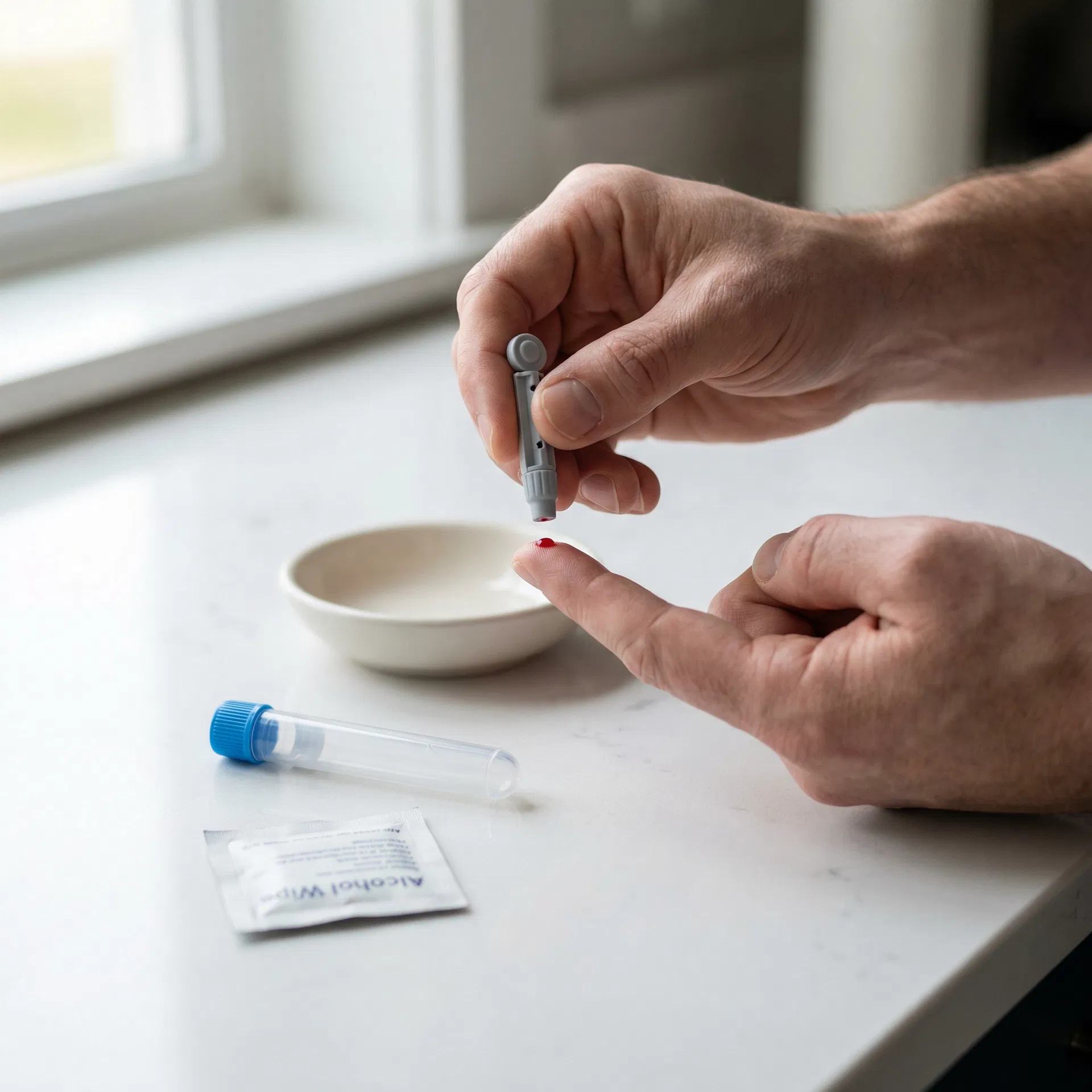
Collect your sample
Follow the simple instructions in your kit — whether it's a finger-prick at home or a venous draw at a partner clinic.
 04
04
Insights delivered
Clear, easy-to-understand results sent to you online with actionable health guidance.
Frequently asked questions
This test measures Total Cholesterol, LDL Cholesterol, Non-HDL Cholesterol, HDL Cholesterol, Total Cholesterol : HDL Ratio. Check the full biomarker list on this page for detailed descriptions.
Check the Special Instructions on this page. General rule: fast 8-12 hours if cholesterol/glucose/insulin included. Most hormone, vitamin, and antibody tests do not require fasting. Morning collection (7-10am) is preferred.
Follow the instructions in your kit. For finger-prick: warm hands, use lancet as directed, fill tube to marked line. For venous: attend a phlebotomy clinic with your lab form. Post same day, avoid Fridays/bank holidays.
Results are typically available within the timeframe shown on this page. You will receive a notification when ready to view online.
You may also be interested in

Female Infertility Check Blood test
A 4-biomarker venous blood panel covering female fertility hormones — ideal for investigating female fertility and reproductive health.
Results estimated in 2 working days
4 biomarkers

Testosterone, Free Testosterone and Oestradiol Blood Test
A 5-biomarker venous blood panel covering hormones and proteins — ideal for investigating low testosterone, libido, energy, or hormone replacement monitoring.
Results estimated in 2 working days
5 biomarkers

Testosterone and Cortisol Blood Test Kit
A 3-biomarker venous blood panel covering hormones — ideal for investigating stress, fatigue, or adrenal function.
Results estimated in 3 working days
3 biomarkers

Allergy Screen (Essential Foods) Blood Test
An essential food allergy panel testing for the most common food allergens responsible for IgE-mediated reactions — covering the key foods that account for the majority of food allergies in both children and adults.
Results estimated in 2 working days
9 biomarkers