
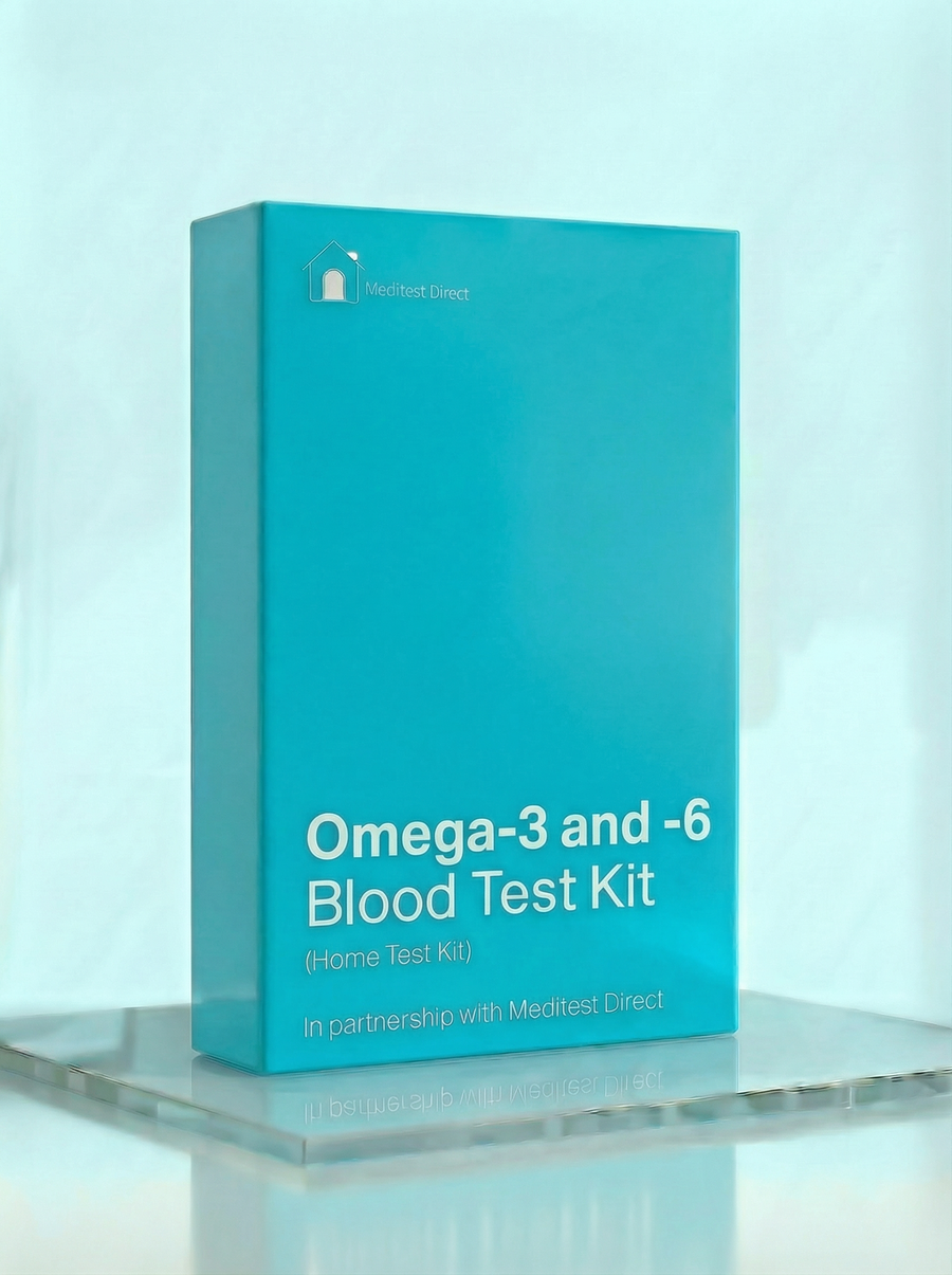
Optimal Health Blood Test Kit
£248 ✓ In Stock
Your sample goes to a UKAS accredited laboratory meeting ISO 15189 standards.
After you receive your order confirmation email, please reply with your date of birth.
How it works
Your testing journey
From order to results in four simple steps. Full transparency on where each step happens and what it costs.

Receive your kit by post
Dispatched same working day if ordered before 3pm. Royal Mail Tracked delivery, typically 1–3 working days. 90% of kits arrive within 24 hours.

Visit a partner clinic
Book a phlebotomy appointment at one of our 365+ UK partner clinics. Take your kit with you — the phlebotomist will collect your sample using the materials provided.
Phlebotomy fee applies (paid at clinic)
Venous blood draw at a clinic
A trained phlebotomist takes a small blood sample from a vein in your arm using the vacutainers provided in your kit. The appointment takes around 10 minutes.

Return by prepaid envelope
Seal your sample in the biohazard bag provided and drop it in any Royal Mail postbox using the prepaid Tracked 24 envelope. Post Monday–Thursday for best results.
Venous Blood Collection Kit
This kit is sent to you and taken to your chosen clinic. The phlebotomist will collect your sample using the materials provided.
- 1Vacutainer blood collection tubes
- 2Needle and butterfly needle
- 3Tourniquet
- 4Alcohol swab
- 5Cotton wool and gauze
- 6Adhesive plaster
- 7Biohazard specimen bag
- 8Prepaid return envelope (Royal Mail Tracked 24)
- 9Laboratory request form
- 10Instructions for the phlebotomist
Total protein measures the combined amount of albumin and globulins in your blood. These proteins perform numerous vital functions including maintaining oncotic pressure (keeping fluid in blood vessels), transporting hormones, vitamins, and medications, contributing to immune defence (immunoglobulins), blood clotting (clotting factors), and enzyme activity. Normal total protein typically ranges from 60-80 g/L. Low total protein can result from reduced production (liver disease, malnutrition), increased loss (kidney disease with proteinuria, protein-losing enteropathy, severe burns), or dilution (fluid overload). Symptoms of low protein include oedema (swelling, particularly in the legs and around the eyes), ascites (abdominal fluid accumulation), and increased susceptibility to infections. High total protein is usually due to elevated globulins rather than albumin. Causes include chronic infections (stimulating immunoglobulin production), chronic inflammatory conditions, dehydration (concentrating proteins), and plasma cell disorders like multiple myeloma. Total protein is most useful when broken down into its components—albumin and globulin—to understand which protein fraction is abnormal. Results outside the normal range may need a follow-up with your GP.
Albumin is the most abundant protein in blood plasma, making up about 55-60% of total protein. It's produced exclusively by the liver at a rate of about 10-15 grams per day. Albumin serves crucial functions: maintaining oncotic pressure (preventing fluid from leaking out of blood vessels into tissues), transporting many substances (hormones, fatty acids, bilirubin, medications), and acting as an antioxidant. Normal albumin levels range from 35-50 g/L. Low albumin (hypoalbuminaemia) can result from reduced production (liver disease, malnutrition, chronic inflammation), increased loss (kidney disease causing proteinuria, protein-losing enteropathy, burns), or dilution. Albumin is a "negative acute phase reactant"—it decreases during inflammation as the liver prioritises production of acute phase proteins instead. Therefore, low albumin during illness doesn't necessarily indicate liver dysfunction or malnutrition. The consequences of low albumin include oedema (fluid accumulation in tissues due to reduced oncotic pressure), impaired drug transport and metabolism, and prolonged wound healing. Albumin levels below 30 g/L are clinically significant and warrant investigation. High albumin is rare and usually indicates dehydration rather than true protein excess. Results outside the normal range may need a follow-up with your GP.
Globulins are a diverse group of proteins that include immunoglobulins (antibodies), transport proteins, clotting factors, and enzymes. They're produced by the liver and the immune system (specifically plasma cells, which produce immunoglobulins). Globulin is calculated by subtracting albumin from total protein. Normal globulin levels typically range from 20-35 g/L. The albumin:globulin ratio (normally above 1) provides additional diagnostic information. Elevated globulins most commonly indicate increased immunoglobulin production. This can occur with chronic infections (HIV, hepatitis B and C, tuberculosis), autoimmune diseases (rheumatoid arthritis, lupus, Sjögren's syndrome), chronic liver disease (particularly cirrhosis), and plasma cell disorders (multiple myeloma, Waldenström's macroglobulinaemia). If globulins are significantly elevated, serum protein electrophoresis can identify which specific proteins are increased. Low globulins can indicate immunodeficiency (reduced antibody production), protein loss, or reduced protein synthesis. Congenital immunodeficiencies, certain medications (including corticosteroids), and malnutrition can cause low globulin levels. The albumin:globulin ratio is reversed (below 1) when globulins exceed albumin—this pattern is seen in cirrhosis, chronic infections, and myeloma. Results outside the normal range may need a follow-up with your GP.
Thyroid Stimulating Hormone is produced by the pituitary gland in the brain and controls the thyroid gland's production of thyroid hormones (T4 and T3). TSH operates in a negative feedback loop: when thyroid hormone levels drop, the pituitary releases more TSH to stimulate the thyroid; when thyroid hormone levels rise, TSH production decreases. This makes TSH the most sensitive marker for thyroid dysfunction—it changes before the thyroid hormones themselves become abnormal. Normal TSH typically ranges from 0.4-4.0 mU/L. High TSH indicates hypothyroidism (underactive thyroid)—the pituitary is "shouting" at a thyroid that isn't responding adequately. Common causes include Hashimoto's thyroiditis (autoimmune thyroid destruction), iodine deficiency, thyroid surgery or radioactive iodine treatment, and certain medications. Symptoms include fatigue, weight gain, cold intolerance, constipation, dry skin, hair loss, and depression. Low TSH indicates hyperthyroidism (overactive thyroid)—the pituitary is responding to excess thyroid hormone by reducing its stimulation signal. Common causes include Graves' disease (autoimmune thyroid stimulation), toxic nodular goitre, thyroiditis, and excessive thyroid hormone replacement. Symptoms include weight loss, rapid heartbeat, anxiety, tremor, heat intolerance, and loose bowels. TSH follows a circadian rhythm (highest early morning), so morning sampling provides the most consistent results. Results outside the normal range may need a follow-up with your GP.
Free T3 (free triiodothyronine) is the metabolically active thyroid hormone. While the thyroid gland produces mostly T4, about 80% of T3 is generated by conversion from T4 in peripheral tissues (liver, kidneys, and other organs). T3 is approximately 3-4 times more potent than T4 and is the hormone that actually acts on cells to regulate metabolism. Only about 0.3% of total T3 circulates in its "free" (unbound) form—the rest is bound to proteins. Free T3 is the biologically active fraction. Normal Free T3 typically ranges from 3.1-6.8 pmol/L. Low Free T3 can indicate hypothyroidism, but more commonly reflects "low T3 syndrome" (euthyroid sick syndrome)—a normal adaptive response to illness, fasting, or stress where the body reduces T4-to-T3 conversion to lower metabolic rate. Low T3 in the context of normal or only slightly elevated TSH during illness usually doesn't indicate thyroid disease and resolves when the underlying condition improves. High Free T3 occurs in hyperthyroidism and is often the earliest hormone change in Graves' disease. Occasionally, T3 toxicosis occurs where Free T3 is elevated while Free T4 remains normal. T3 is also elevated with excessive intake of T3-containing thyroid hormone preparations (liothyronine or desiccated thyroid extract). Results outside the normal range may need a follow-up with your GP.
Free T4 (free thyroxine) is the main hormone produced by the thyroid gland. About 99.97% of T4 in the blood is bound to transport proteins (thyroxine-binding globulin, albumin, and transthyretin); only the tiny "free" fraction (about 0.03%) is biologically available. Free T4 serves primarily as a reservoir that's converted to the more active T3 in target tissues. Measuring Free T4 (rather than Total T4) avoids interference from protein-binding variations. Normal Free T4 typically ranges from 12-22 pmol/L. Low Free T4 with high TSH confirms primary hypothyroidism—the thyroid gland is failing to produce adequate hormone despite maximal stimulation. The most common cause is Hashimoto's thyroiditis. Treatment with levothyroxine (synthetic T4) replaces the deficient hormone. Low Free T4 with low or normal TSH suggests secondary (pituitary) or tertiary (hypothalamic) hypothyroidism—rare conditions where the problem lies in the control systems rather than the thyroid itself. High Free T4 with low TSH confirms hyperthyroidism—excess thyroid hormone production is suppressing pituitary TSH. Common causes include Graves' disease and toxic nodular goitre. High Free T4 can also result from excessive thyroid hormone medication (overreplacement). In rare cases, high Free T4 with non-suppressed TSH can indicate a TSH-producing pituitary adenoma or thyroid hormone resistance syndrome. Results outside the normal range may need a follow-up with your GP.
Serum iron measures the amount of iron circulating in your blood, bound to the transport protein transferrin. Iron is essential for oxygen transport (as part of haemoglobin in red blood cells), energy production (as part of enzymes in the mitochondria), DNA synthesis, and immune function. The body tightly regulates iron because both deficiency and excess are harmful. Normal serum iron typically ranges from 10-30 µmol/L, though it varies significantly throughout the day. Serum iron alone is not a reliable indicator of iron status because it fluctuates considerably—it's highest in the morning and can vary by 30-40% within the same day. Recent iron-rich meals, iron supplements, and even the time of blood collection significantly affect results. For this reason, serum iron is always interpreted alongside other iron markers, particularly ferritin, TIBC, and transferrin saturation. Low serum iron can indicate iron deficiency but can also occur in chronic disease (anaemia of chronic disease) where iron is sequestered and not released into circulation. High serum iron may suggest iron overload conditions (haemochromatosis), recent iron supplementation, or haemolysis (breakdown of red blood cells releasing their iron content). Results outside the normal range should be interpreted alongside other iron markers and may need a follow-up with your GP.
Total Iron Binding Capacity measures the maximum amount of iron that transferrin proteins in your blood can carry. Transferrin is the transport protein that picks up iron from the gut (after absorption) and from storage sites, and delivers it to cells that need it, particularly the bone marrow for red blood cell production. TIBC essentially reflects the amount of transferrin available to bind iron. Normal TIBC typically ranges from 45-80 µmol/L. TIBC rises when the body needs more iron and falls when there's plenty. In iron deficiency, the liver produces more transferrin to maximise iron capture and transport, so TIBC increases. In iron overload or chronic inflammation, less transferrin is needed, so TIBC decreases. This inverse relationship makes TIBC a useful complement to serum iron for understanding iron status. The combination of serum iron and TIBC allows calculation of transferrin saturation (serum iron ÷ TIBC × 100), which indicates what percentage of iron-carrying capacity is being used. Low transferrin saturation with high TIBC suggests iron deficiency. High transferrin saturation with low/normal TIBC suggests iron overload. Results outside the normal range may need a follow-up with your GP.
Transferrin saturation (TSAT) is the percentage of transferrin that is currently bound to iron, calculated by dividing serum iron by TIBC and multiplying by 100. It indicates how much of your iron transport capacity is being utilised. Normal transferrin saturation typically ranges from 20-45%, meaning that normally about a quarter to half of your transferrin molecules are carrying iron at any given time. Low transferrin saturation (below 20%) suggests iron deficiency—there's not enough iron to occupy the available transport capacity. This is one of the earlier indicators of iron deficiency, often becoming abnormal before haemoglobin drops into the anaemic range. In iron deficiency anaemia, transferrin saturation is typically below 15%. High transferrin saturation (above 45%) suggests iron overload—more iron is circulating than normal. In hereditary haemochromatosis, transferrin saturation is often above 50-60% and is one of the earliest screening markers for this condition. Very high saturation (above 80-90%) means almost all transferrin is iron-bound, and "free" iron may begin circulating, which can cause tissue damage. Persistently elevated transferrin saturation warrants further investigation, including genetic testing for haemochromatosis. Results outside the normal range may need a follow-up with your GP.
Ferritin is the primary iron storage protein in the body. A small amount of ferritin circulates in the blood, and measuring serum ferritin provides a window into total body iron stores. Each ferritin molecule can store up to 4,500 iron atoms, making it an efficient storage system. Normal ferritin levels vary by sex: typically 30-400 µg/L for men and 15-150 µg/L for premenopausal women (with higher upper limits after menopause). Low ferritin is the most specific marker for iron deficiency. Ferritin below 30 µg/L strongly suggests depleted iron stores, and levels below 15 µg/L are diagnostic of iron deficiency. Symptoms of low ferritin include fatigue, weakness, poor concentration, hair loss, restless legs, and if severe enough to cause anaemia, shortness of breath and pallor. Iron deficiency is the most common nutritional deficiency worldwide, particularly affecting women of reproductive age due to menstrual blood loss. High ferritin can indicate iron overload (haemochromatosis), but ferritin is also an acute phase reactant—it rises with inflammation, infection, liver disease, and some malignancies independently of iron stores. This is why hs-CRP is included in this panel: if both ferritin and CRP are elevated, the raised ferritin may reflect inflammation rather than true iron overload. Persistently elevated ferritin with normal CRP warrants investigation for haemochromatosis or other causes of iron overload. Results outside the normal range may need a follow-up with your GP.
Unsaturated Iron Binding Capacity measures the reserve capacity of transferrin to bind additional iron—essentially, how much "room" remains on transferrin molecules that aren't currently carrying iron. UIBC represents the portion of TIBC that is not occupied by iron. Mathematically, TIBC = Serum Iron + UIBC. Normal UIBC typically ranges from 25-60 µmol/L. In iron deficiency, UIBC increases because transferrin molecules have plenty of empty binding sites waiting for iron that isn't available. The body has upregulated transferrin production to capture as much iron as possible, but there's not enough iron to fill these transport proteins. In iron overload, UIBC decreases because most transferrin binding sites are already occupied by iron—there's little reserve capacity left. Very low UIBC with high serum iron and high transferrin saturation is the classic pattern of haemochromatosis or other iron overload states. UIBC provides complementary information to the other iron markers and helps confirm patterns of deficiency or excess. Results outside the normal range may need a follow-up with your GP.
Bilirubin is a yellow pigment produced when the liver breaks down old red blood cells. Haemoglobin from these cells is converted to bilirubin, which the liver processes (conjugates) and excretes into bile. From there, bilirubin travels to the intestines and gives stool its characteristic brown colour. Normal total bilirubin levels are typically below 21 µmol/L. Elevated bilirubin causes jaundice—yellowing of the skin and whites of the eyes. This can result from increased production (excessive breakdown of red blood cells, as in haemolytic anaemia), impaired liver processing (hepatitis, cirrhosis, medications), or blocked excretion (gallstones, bile duct obstruction, pancreatic cancer). The pattern of elevation (conjugated vs unconjugated bilirubin) helps identify the cause. Gilbert's syndrome is a common, benign inherited condition affecting about 5% of the population where bilirubin levels are mildly elevated (typically 20-50 µmol/L) due to reduced liver enzyme activity. It causes no symptoms other than occasional mild jaundice, particularly during fasting, stress, or illness. If your bilirubin is mildly elevated with normal liver enzymes and you feel well, Gilbert's syndrome is the likely explanation. Results significantly outside the normal range or associated with other abnormal liver markers may need a follow-up with your GP.
Alkaline phosphatase is an enzyme found in many tissues, but mainly in liver, bone, kidneys, and intestines. In liver, ALP is concentrated in the cells lining the bile ducts. In bone, it's produced by osteoblasts (bone-building cells). Because ALP comes from multiple sources, elevated levels require interpretation alongside other markers to determine the origin. Normal ALP typically ranges from 30-130 U/L in adults. Elevated ALP from liver sources typically indicates bile duct obstruction or cholestatic (bile flow) problems—gallstones, primary biliary cholangitis, primary sclerosing cholangitis, or medications causing cholestasis. In these cases, GGT is usually also elevated, helping confirm the liver origin. Isolated ALP elevation with normal GGT suggests a bone source. Elevated ALP from bone sources occurs during active bone formation or turnover—fracture healing, Paget's disease, bone metastases, vitamin D deficiency with secondary hyperparathyroidism, or during normal growth in children and adolescents. Physiologically elevated ALP also occurs in the third trimester of pregnancy (placental ALP). Low ALP is rare but can occur in hypothyroidism, anaemia, or certain rare genetic conditions. Results outside the normal range may need a follow-up with your GP.
Alanine aminotransferase is an enzyme found predominantly in liver cells (hepatocytes), with smaller amounts in kidneys, heart, and muscles. Because it's concentrated in the liver, ALT is considered the most liver-specific of the common liver enzymes. When liver cells are damaged or inflamed, they release ALT into the bloodstream, causing levels to rise. Normal ALT is typically below 40 U/L, though optimal levels are likely lower. Elevated ALT indicates hepatocyte injury. The most common cause in the UK is non-alcoholic fatty liver disease (NAFLD), associated with obesity, insulin resistance, and metabolic syndrome. Other causes include alcohol-related liver disease, viral hepatitis (B and C), medications (including paracetamol, statins, and many others), autoimmune hepatitis, and rarer conditions. Very high ALT (more than 10 times normal) suggests acute liver injury from viral hepatitis, drug toxicity, or ischaemia. The degree of ALT elevation doesn't always correlate with the severity of underlying liver disease—some serious conditions like cirrhosis may have only mildly elevated or even normal ALT. Mildly elevated ALT should prompt investigation of lifestyle factors (weight, alcohol, medications) and consideration of further testing including liver ultrasound. Results outside the normal range may need a follow-up with your GP.
Gamma-glutamyl transferase is an enzyme found in liver, bile ducts, kidney, pancreas, and other tissues. In clinical practice, GGT is primarily used as a marker of liver and bile duct health, and notably as a sensitive indicator of alcohol consumption. Normal GGT is typically below 50 U/L in women and below 60 U/L in men, though some laboratories use different reference ranges. GGT is induced by alcohol and many medications, meaning these substances cause the liver to produce more GGT enzyme. Regular alcohol consumption—even at moderate levels—commonly elevates GGT, and it's estimated that GGT is elevated in approximately 75% of heavy drinkers. GGT typically falls within 2-6 weeks of abstinence, making it useful for monitoring alcohol reduction. GGT is also elevated by many medications including anticonvulsants, certain antibiotics, and enzyme-inducing drugs. Elevated GGT alongside elevated ALP suggests bile duct pathology (since both are concentrated in bile duct cells), while elevated GGT with elevated ALT suggests hepatocyte damage affecting both parenchymal and bile duct cells. GGT alone (without other liver enzyme elevation) is often due to alcohol or medications and may not indicate significant liver disease. However, persistently elevated GGT has been associated with increased cardiovascular and all-cause mortality in some studies, independent of alcohol intake. Results outside the normal range may need a follow-up with your GP.
Urea is a waste product formed in the liver when protein is broken down. Amino acids from dietary protein and from the body's own tissue turnover are metabolised, producing ammonia as a byproduct. The liver converts this toxic ammonia into urea, which travels via the bloodstream to the kidneys for excretion in urine. Measuring blood urea helps assess both kidney function (the kidneys' ability to excrete urea) and protein metabolism. Normal urea typically ranges from 2.5-7.8 mmol/L. Elevated urea can indicate reduced kidney function (the kidneys aren't excreting urea efficiently), but it's less specific than creatinine because urea levels are also affected by protein intake, dehydration, gastrointestinal bleeding (digested blood acts as a protein load), certain medications, and catabolic states (where the body is breaking down its own tissue). The urea:creatinine ratio can help distinguish between different causes of elevated urea. Low urea may occur with low protein intake, severe liver disease (reduced urea production), or overhydration. In isolation, low urea is rarely clinically significant. Urea is most useful when interpreted alongside creatinine and eGFR for a complete picture of kidney function. Results outside the normal range may need a follow-up with your GP.
Creatinine is a waste product generated from the normal breakdown of creatine phosphate in muscle tissue. Because it's produced at a relatively constant rate (proportional to muscle mass) and is filtered almost exclusively by the kidneys, creatinine is one of the most reliable markers for assessing kidney function. Normal creatinine levels typically range from 60-110 µmol/L in men and 45-90 µmol/L in women, though this varies with muscle mass. Elevated creatinine indicates reduced kidney filtration. The kidneys normally filter creatinine from the blood and excrete it in urine; when kidney function declines, creatinine accumulates in the blood. However, creatinine may not rise above the normal range until kidney function has declined by approximately 50%, making it a relatively late marker of kidney disease. This is why eGFR (which adjusts creatinine for age, sex, and ethnicity) is more sensitive for detecting early kidney impairment. Creatinine can also be elevated temporarily after intense exercise, with high protein/meat intake (particularly creatine supplements), or with certain medications that affect kidney filtration or creatinine secretion. Very low creatinine may be seen in people with low muscle mass, malnutrition, or liver disease. Results outside the normal range may need a follow-up with your GP.
The estimated Glomerular Filtration Rate calculates how much blood your kidneys filter per minute, expressed in mL/min/1.73m². It's derived from your creatinine level using the CKD-EPI formula, which adjusts for age and sex (and ethnicity in some versions) to provide a more accurate estimate of kidney function than creatinine alone. A normal eGFR is above 90 mL/min/1.73m². eGFR is used to stage chronic kidney disease (CKD): Stage 1 (≥90 with other evidence of kidney damage), Stage 2 (60-89), Stage 3a (45-59), Stage 3b (30-44), Stage 4 (15-29), and Stage 5 (below 15 or on dialysis). Importantly, eGFR must be persistently reduced for more than 3 months to diagnose CKD—a single low reading may reflect temporary factors like dehydration or acute illness. Declining eGFR over time is more concerning than a single static measurement. Regular monitoring can track kidney function trajectory. Factors that accelerate kidney decline include poorly controlled diabetes, hypertension, recurrent kidney infections, certain medications (especially NSAIDs and some antibiotics), and obstruction. Early detection through eGFR monitoring allows interventions to slow progression. Results outside the normal range may need a follow-up with your GP.
Sodium is the primary electrolyte in extracellular fluid (the fluid outside cells, including blood plasma). It plays essential roles in maintaining fluid balance, blood pressure, nerve impulse transmission, and muscle contraction. The kidneys are the primary regulators of sodium balance, adjusting sodium excretion based on intake and the body's needs. Normal serum sodium ranges from 136-145 mmol/L. Low sodium (hyponatraemia, below 136 mmol/L) is relatively common and usually reflects water balance rather than sodium deficiency—it typically means there's too much water relative to sodium. Causes include excessive fluid intake, certain medications (particularly diuretics, antidepressants, and some blood pressure medications), heart failure, liver cirrhosis, kidney disease, and the syndrome of inappropriate ADH secretion (SIADH). Mild hyponatraemia may cause no symptoms, but severe cases can cause confusion, seizures, and coma. High sodium (hypernatraemia, above 145 mmol/L) usually indicates dehydration—water loss exceeding sodium loss. It can occur with inadequate fluid intake (particularly in elderly people with reduced thirst sensation), excessive sweating, diarrhoea, diabetes insipidus, or certain medications. Symptoms include thirst, confusion, and in severe cases, seizures. Results outside the normal range may need a follow-up with your GP.
Folate (vitamin B9) is essential for DNA synthesis, cell division, and amino acid metabolism. It's particularly crucial during periods of rapid cell division—pregnancy, infancy, and growth. Folate works closely with vitamin B12 in one-carbon metabolism and red blood cell production. Good dietary sources include leafy green vegetables, legumes, citrus fruits, and fortified foods. The body stores relatively little folate, so regular intake is important. Normal serum folate typically ranges from 7-45 nmol/L. Folate deficiency can result from inadequate dietary intake (poor diet, alcohol excess), increased requirements (pregnancy, haemolytic anaemia, cancer), malabsorption (coeliac disease, inflammatory bowel disease), or medications that interfere with folate metabolism (methotrexate, phenytoin, trimethoprim). Deficiency causes megaloblastic anaemia (large, immature red cells), similar to B12 deficiency. Symptoms include fatigue, weakness, shortness of breath, and neurological symptoms (though neurological effects are more prominent with B12 deficiency). Serum folate reflects recent dietary intake over the past few days—a single high-folate meal can temporarily normalise levels. Red cell folate (not included in this panel) better reflects long-term status over the preceding 2-3 months. Low serum folate should prompt dietary review and consideration of causes of deficiency. In pregnant women, folate deficiency increases the risk of neural tube defects in the developing baby—this is why folic acid supplementation is recommended before conception and during early pregnancy. Results outside the normal range may need a follow-up with your GP.
Active B12 (holotranscobalamin) measures the biologically available form of vitamin B12. Total B12 measures all B12 in the blood, but 70-90% is bound to haptocorrin and is not available to cells. Active B12 specifically measures B12 bound to transcobalamin II—the transport protein that delivers B12 into cells. This makes Active B12 a more sensitive and specific marker for true B12 status than Total B12, particularly for detecting early deficiency. Normal Active B12 is typically above 25 pmol/L. B12 is essential for DNA synthesis, neurological function, and red blood cell production. It's found exclusively in animal products (meat, fish, eggs, dairy), making deficiency common in vegans and vegetarians who don't supplement. B12 requires intrinsic factor (produced by stomach cells) for absorption in the terminal ileum. Deficiency can result from inadequate intake (vegan diet), malabsorption (pernicious anaemia destroying intrinsic factor-producing cells, coeliac disease, Crohn's disease, gastric surgery), or certain medications (metformin, proton pump inhibitors). B12 deficiency causes megaloblastic anaemia (large, immature red cells similar to folate deficiency) and, importantly, neurological damage that can be irreversible if not treated promptly. Neurological symptoms include numbness and tingling in hands and feet (peripheral neuropathy), balance problems, cognitive changes, depression, and in severe cases, spinal cord degeneration. Active B12 below 25 pmol/L is concerning for deficiency; levels 25-70 pmol/L are indeterminate and may warrant further testing (MMA, homocysteine). Results outside the normal range may need a follow-up with your GP.
Haemoglobin is the iron-containing protein in red blood cells that carries oxygen from the lungs to tissues and returns carbon dioxide to the lungs for exhalation. Each haemoglobin molecule contains four haem groups, each with an iron atom that can bind one oxygen molecule. Haemoglobin gives blood its red colour—bright red when oxygenated, darker when deoxygenated. Normal haemoglobin is typically 130-170 g/L in men and 120-150 g/L in women. Low haemoglobin (anaemia) means reduced oxygen-carrying capacity. Symptoms include fatigue, weakness, shortness of breath (particularly on exertion), pallor, dizziness, and rapid heartbeat as the body compensates for reduced oxygen delivery. Anaemia has many causes: iron deficiency (most common), vitamin B12 or folate deficiency, chronic disease, blood loss, bone marrow disorders, haemolysis (red cell destruction), and inherited conditions like thalassaemia. High haemoglobin (polycythaemia) increases blood viscosity and clotting risk. It can be primary (polycythaemia vera, a bone marrow disorder) or secondary (response to chronic hypoxia from lung disease, sleep apnoea, living at high altitude, or smoking; or inappropriate erythropoietin production from certain tumours). Dehydration can also artificially elevate haemoglobin by concentrating the blood. Results outside the normal range may need a follow-up with your GP.
Haematocrit (also called packed cell volume or PCV) measures the percentage of blood volume occupied by red blood cells. When blood is centrifuged, red cells settle at the bottom, and haematocrit represents this packed red cell layer as a proportion of total blood volume. Normal haematocrit is typically 40-52% in men and 36-48% in women. Haematocrit correlates closely with haemoglobin—both reflect red cell mass. Low haematocrit indicates anaemia or haemodilution (excess fluid diluting the blood). The causes mirror those for low haemoglobin: iron deficiency, vitamin deficiencies, chronic disease, blood loss, bone marrow failure, and haemolysis. Overhydration (from excessive IV fluids or certain medical conditions) can lower haematocrit without true reduction in red cell mass. High haematocrit increases blood viscosity, making the blood "thicker" and harder to pump. This increases the risk of blood clots, stroke, and heart attack. Causes include dehydration (concentrating red cells), polycythaemia vera, chronic hypoxia (lung disease, sleep apnoea, high altitude, smoking), and erythropoietin-producing tumours. Haematocrit above 50% in women or 52% in men warrants investigation. Results outside the normal range may need a follow-up with your GP.
Red cell count measures the number of red blood cells (erythrocytes) per litre of blood. Red blood cells are the most numerous cells in blood—approximately 4.5-5.5 million per microlitre in men and 4.0-5.0 million per microlitre in women. They're produced in the bone marrow, live for approximately 120 days, and are then removed by the spleen. Red cell count, along with haemoglobin and haematocrit, helps assess overall red cell mass. Low red cell count occurs in anaemia from any cause—reduced production (nutritional deficiencies, bone marrow disorders, chronic disease), increased destruction (haemolytic anaemias), or blood loss. The red cell count should be interpreted alongside MCV (cell size) to classify the type of anaemia: microcytic (small cells, often iron deficiency), normocytic (normal size, often chronic disease), or macrocytic (large cells, often B12/folate deficiency). High red cell count (erythrocytosis) can be appropriate (living at high altitude, chronic lung disease) or inappropriate (polycythaemia vera, erythropoietin-producing tumours). It's also seen with dehydration, which concentrates red cells. High red cell counts increase blood viscosity and thrombosis risk. Results outside the normal range may need a follow-up with your GP.
Mean Corpuscular Volume measures the average size of your red blood cells in femtolitres (fL). It's calculated from the haematocrit and red cell count. MCV is one of the most useful indices for classifying and diagnosing anaemia. Normal MCV typically ranges from 80-100 fL. Low MCV (microcytosis, below 80 fL) indicates smaller-than-normal red cells. The most common cause is iron deficiency—without adequate iron, the bone marrow produces smaller cells with less haemoglobin. Other causes include thalassaemia (inherited haemoglobin disorders), anaemia of chronic disease, lead poisoning, and sideroblastic anaemia. Iron deficiency and thalassaemia trait can both cause microcytosis with similar MCV, but other indices and iron studies help distinguish them. High MCV (macrocytosis, above 100 fL) indicates larger-than-normal red cells. Common causes include vitamin B12 deficiency, folate deficiency, alcohol excess, liver disease, hypothyroidism, certain medications (methotrexate, anticonvulsants, azathioprine), and bone marrow disorders (myelodysplasia). B12 and folate are required for DNA synthesis; their deficiency impairs cell division, leading to larger, immature red cells. Alcohol directly affects red cell membrane composition and bone marrow function. Results outside the normal range may need a follow-up with your GP.
Mean Corpuscular Haemoglobin measures the average amount of haemoglobin in each red blood cell, expressed in picograms (pg). It's calculated from haemoglobin concentration and red cell count. MCH reflects how much oxygen-carrying capacity each red cell has. Normal MCH typically ranges from 27-32 pg. Low MCH (hypochromic cells) indicates less haemoglobin per cell than normal. This typically parallels low MCV, as smaller cells contain less haemoglobin. Iron deficiency is the most common cause—without sufficient iron, haemoglobin synthesis is impaired, producing pale, small cells with reduced MCH. Thalassaemia and chronic disease anaemia also cause low MCH. High MCH typically occurs alongside high MCV (macrocytosis), as larger cells contain more haemoglobin. Causes include B12 deficiency, folate deficiency, and other causes of macrocytosis. MCH and MCHC provide complementary information to MCV when characterising red cell abnormalities. Results outside the normal range may need a follow-up with your GP.
Mean Corpuscular Haemoglobin Concentration measures the average concentration of haemoglobin within red blood cells, expressed in grams per litre of red cells (g/L). Unlike MCH (total haemoglobin per cell), MCHC reflects how densely packed the haemoglobin is within each cell. Normal MCHC typically ranges from 320-360 g/L. Low MCHC indicates hypochromic cells—red cells that are paler than normal because they contain dilute haemoglobin. This is characteristic of iron deficiency, where there's insufficient iron to produce adequate haemoglobin, and thalassaemia, where haemoglobin synthesis is impaired. Cells in these conditions are not only small (low MCV) but also pale (low MCHC). High MCHC is relatively uncommon and may indicate hereditary spherocytosis (where abnormal cell membranes cause red cells to become sphere-shaped and concentrated), severe dehydration, or laboratory artefact. MCHC above 370 g/L should prompt consideration of spherocytosis, particularly if there's a family history of anaemia, jaundice, or splenectomy. Results outside the normal range may need a follow-up with your GP.
Red Cell Distribution Width measures the variation in red blood cell sizes—essentially, how much your red cells differ from each other in volume. It's expressed as a percentage coefficient of variation. In healthy individuals, red cells are fairly uniform in size, giving a normal RDW of 11.5-15%. A high RDW indicates increased variation (anisocytosis)—cells of many different sizes. Elevated RDW is clinically useful for distinguishing causes of anaemia. In iron deficiency anaemia, RDW is typically elevated because the bone marrow produces progressively smaller cells as iron stores deplete, while older normal-sized cells remain in circulation—creating size variation. In thalassaemia trait, which also causes microcytosis, RDW is typically normal because all cells are uniformly small. This distinction helps differentiate iron deficiency from thalassaemia when both present with low MCV. RDW is also elevated in mixed nutritional deficiencies (iron plus B12 or folate), recent blood transfusion (donor and recipient cells differ), recovering anaemia (new normal-sized cells mixing with old abnormal cells), and certain bone marrow disorders. Recent research has also associated elevated RDW with cardiovascular disease and mortality risk, possibly reflecting chronic inflammation or nutritional inadequacy. Results outside the normal range may need a follow-up with your GP.
White cell count measures the total number of white blood cells (leukocytes) in your blood. White cells are the soldiers of your immune system—they defend against infection, remove debris, and are involved in allergic and inflammatory responses. There are five main types: neutrophils, lymphocytes, monocytes, eosinophils, and basophils, each with different functions. Normal WCC typically ranges from 4.0-11.0 × 10⁹/L. High white cell count (leukocytosis) most commonly indicates infection—the bone marrow ramps up production to fight invading pathogens. Other causes include inflammation, tissue injury, stress, medications (particularly corticosteroids), smoking, and rarely, leukaemia (cancer of white blood cells). The differential count (which types of white cells are elevated) helps identify the cause: bacterial infections typically raise neutrophils, viral infections may raise lymphocytes, and allergic conditions raise eosinophils. Low white cell count (leukopenia) increases infection risk because you have fewer immune cells to fight pathogens. Causes include viral infections (including HIV), bone marrow disorders, certain medications (chemotherapy, immunosuppressants), autoimmune conditions attacking white cells, and severe infections overwhelming the immune system. Neutropenia (low neutrophils specifically) is the most clinically significant because neutrophils are the primary defence against bacterial infections. Results outside the normal range may need a follow-up with your GP.
Neutrophils are the most abundant white blood cells, making up 50-70% of the total. They're the first responders to bacterial infections—they migrate to infection sites, engulf (phagocytose) bacteria, and release antimicrobial substances. Neutrophils live only 5-90 hours in circulation before migrating to tissues or dying. The bone marrow maintains a large reserve and can rapidly increase production during infection. Normal neutrophil count is typically 2.0-7.5 × 10⁹/L. High neutrophils (neutrophilia) most commonly indicate bacterial infection. Other causes include inflammation (appendicitis, pancreatitis, inflammatory bowel disease), tissue damage (heart attack, surgery, burns), medications (corticosteroids, lithium), smoking, stress, and rarely chronic myeloid leukaemia. The neutrophil count often rises rapidly during acute bacterial infections and may be accompanied by "left shift"—the appearance of immature neutrophils (bands) indicating accelerated production. Low neutrophils (neutropenia) significantly increases infection risk. Causes include viral infections (which can temporarily suppress neutrophil production), bone marrow disorders, chemotherapy, certain medications (carbimazole, clozapine, NSAIDs), autoimmune neutropenia, and some ethnic variations (benign ethnic neutropenia, common in people of African descent). Neutrophil counts below 1.0 × 10⁹/L are concerning; below 0.5 × 10⁹/L, the risk of serious bacterial infection increases substantially. Results outside the normal range may need a follow-up with your GP.
Lymphocytes are the cells of adaptive immunity—they recognise specific pathogens and mount targeted immune responses. There are three main types: B lymphocytes (produce antibodies), T lymphocytes (coordinate immune responses and directly kill infected cells), and natural killer cells (destroy virus-infected and cancer cells). Lymphocytes constitute 20-40% of white blood cells. Unlike neutrophils, many lymphocytes are long-lived, retaining immune memory for years or decades. Normal lymphocyte count is typically 1.0-4.0 × 10⁹/L. High lymphocytes (lymphocytosis) commonly occur with viral infections—the lymphocyte response to viruses contrasts with the neutrophil response to bacteria. Infections causing lymphocytosis include Epstein-Barr virus (infectious mononucleosis), cytomegalovirus, hepatitis, HIV, and pertussis. Chronic lymphocytosis may indicate chronic lymphocytic leukaemia (CLL), the most common leukaemia in adults, which typically presents with progressively rising lymphocyte counts in older individuals. Low lymphocytes (lymphopenia) can indicate viral infections (paradoxically, some viruses like HIV destroy lymphocytes), immunodeficiency, corticosteroid use, chemotherapy, radiation therapy, autoimmune diseases like lupus, and severe acute illness. Lymphopenia reduces the body's ability to mount specific immune responses. Low lymphocytes during acute infection may indicate overwhelming infection or poor prognosis. Results outside the normal range may need a follow-up with your GP.
Monocytes are the largest white blood cells and comprise about 2-8% of the total. They circulate in blood for 1-3 days before migrating into tissues, where they mature into macrophages or dendritic cells. Macrophages are powerful phagocytes—they engulf and destroy pathogens, dead cells, and debris. They also present antigens to lymphocytes, bridging innate and adaptive immunity. Dendritic cells are specialised antigen-presenting cells crucial for initiating immune responses. Normal monocyte count is typically 0.2-0.8 × 10⁹/L. High monocytes (monocytosis) can indicate chronic infections (tuberculosis, subacute bacterial endocarditis, brucellosis), inflammatory conditions (inflammatory bowel disease, rheumatoid arthritis), recovery from acute infection (monocytes increase as neutrophils decline), and certain malignancies (chronic myelomonocytic leukaemia, Hodgkin lymphoma). Monocytosis often accompanies chronic rather than acute processes. Low monocytes (monocytopenia) are relatively uncommon. Causes include acute infections (which may temporarily deplete monocytes), bone marrow failure, hairy cell leukaemia, and corticosteroid therapy. Isolated low monocytes are rarely clinically significant unless severe. The monocyte count is most useful in context—persistent monocytosis warrants investigation, while mild fluctuations are usually not concerning. Results outside the normal range may need a follow-up with your GP.
Eosinophils are white blood cells with distinctive granules that stain red-orange with eosin dye. They comprise only 1-4% of circulating white cells but are concentrated in tissues, particularly the gastrointestinal tract, respiratory tract, and skin. Eosinophils evolved primarily to combat parasitic infections—their granules contain proteins toxic to parasites. They also play important roles in allergic reactions and inflammation, releasing mediators that contribute to symptoms of asthma, eczema, and allergies. Normal eosinophil count is typically 0.04-0.4 × 10⁹/L. High eosinophils (eosinophilia) most commonly indicate allergic conditions (asthma, hay fever, eczema, food allergies, drug allergies) or parasitic infections (particularly those with tissue invasion, like roundworms and hookworms). Other causes include autoimmune diseases (eosinophilic granulomatosis with polyangiitis, previously called Churg-Strauss syndrome), certain cancers (Hodgkin lymphoma, some leukaemias), adrenal insufficiency, and idiopathic hypereosinophilic syndrome. In developed countries where parasites are uncommon, allergic disease is the most frequent cause of eosinophilia. However, in someone with recent travel to endemic areas or immigrant populations, parasitic infection should be considered. Very high eosinophil counts (above 1.5 × 10⁹/L) can cause organ damage from eosinophil infiltration and toxic granule release—this is called hypereosinophilic syndrome and requires investigation and treatment. Results outside the normal range may need a follow-up with your GP.
Basophils are the rarest white blood cells, comprising less than 1% of the total. Their granules stain dark blue with basic dyes and contain histamine, heparin, and other inflammatory mediators. Basophils are closely related to tissue mast cells and play similar roles in allergic reactions and inflammation. When activated by allergens (via IgE antibodies on their surface), basophils release their granules (degranulation), contributing to allergy symptoms. Normal basophil count is typically 0.0-0.1 × 10⁹/L. High basophils (basophilia) are uncommon but can indicate allergic reactions, chronic inflammatory conditions (inflammatory bowel disease, rheumatoid arthritis), infections, hypothyroidism, and certain myeloproliferative disorders (chronic myeloid leukaemia, polycythaemia vera, myelofibrosis). Persistent basophilia, particularly if marked, may be an early indicator of myeloproliferative disease and warrants further investigation. Low basophils (basopenia) are difficult to detect reliably given that normal counts are already very low. Causes include acute allergic reactions (basophils degranulate and leave circulation), hyperthyroidism, acute infection, and corticosteroid therapy. Clinically, basophil counts are most useful when persistently elevated, suggesting myeloproliferative disorder or chronic allergic/inflammatory conditions. Isolated low or normal basophils are rarely clinically significant. Results outside the normal range may need a follow-up with your GP.
High-sensitivity C-reactive protein (hs-CRP) is a protein produced by the liver in response to inflammation anywhere in the body. The "high-sensitivity" assay can detect very low levels of CRP, making it useful for assessing chronic low-grade inflammation rather than just acute infections or injuries. CRP is part of the innate immune system and rises rapidly (within hours) in response to inflammatory stimuli, making it a sensitive but non-specific marker. In the context of cardiovascular risk assessment, hs-CRP levels below 1 mg/L are considered low risk, 1-3 mg/L moderate risk, and above 3 mg/L higher risk. Chronic low-grade inflammation, reflected by mildly elevated hs-CRP, is associated with atherosclerosis, metabolic syndrome, and increased cardiovascular events. However, hs-CRP is influenced by many factors including obesity, smoking, gum disease, sleep apnoea, and chronic infections. Levels above 10 mg/L typically indicate acute infection or significant inflammation rather than chronic low-grade inflammation—in this case, the test should be repeated once the acute issue has resolved. hs-CRP is also important for interpreting ferritin levels, as ferritin is an acute phase reactant that rises with inflammation independently of iron stores. If both hs-CRP and ferritin are elevated, the ferritin may be elevated due to inflammation rather than iron overload. Results outside the normal range may need a follow-up with your GP.
Platelets (thrombocytes) are small cell fragments produced in the bone marrow that play a crucial role in blood clotting. When you cut yourself or damage a blood vessel, platelets rush to the site, stick together, and form a plug that helps stop bleeding. They also release chemicals that activate the clotting cascade, leading to the formation of a stable fibrin clot. The normal platelet count ranges from 150,000 to 400,000 per microlitre of blood. Low platelet counts (thrombocytopenia) can result from reduced production (bone marrow disorders, certain medications, viral infections), increased destruction (autoimmune conditions like ITP, some medications), or increased consumption (severe infections, DIC). Symptoms of low platelets include easy bruising, prolonged bleeding from cuts, petechiae (small red spots under the skin), and in severe cases, spontaneous bleeding. High platelet counts (thrombocytosis) can be reactive (responding to infection, inflammation, iron deficiency, or after surgery) or primary (due to bone marrow disorders like essential thrombocythemia). Elevated platelets can increase the risk of blood clots (thrombosis). Results outside the normal range may need a follow-up with your GP.
Mean Platelet Volume measures the average size of your platelets in femtolitres (fL). Platelet size matters because it reflects platelet age and activity—younger platelets freshly released from the bone marrow are typically larger and more metabolically active than older platelets. Normal MPV typically ranges from 7.5 to 11.5 fL, though reference ranges vary between laboratories. A high MPV suggests increased platelet production and turnover. This can occur when platelets are being consumed or destroyed faster than normal (such as in immune thrombocytopenia), prompting the bone marrow to release younger, larger platelets. High MPV has also been associated with increased cardiovascular risk in some studies, as larger platelets are more reactive and may contribute to clot formation. A low MPV may indicate reduced bone marrow production or older platelet populations. It can be seen in certain bone marrow disorders, some inflammatory conditions, and during chemotherapy. MPV is most useful when interpreted alongside the platelet count—for example, low platelets with high MPV suggests the bone marrow is trying to compensate for platelet destruction, while low platelets with low MPV might suggest a production problem. Results outside the normal range may need a follow-up with your GP.
This test is for screening and information only — it is not a medical diagnosis or professional advice. Please have your results reviewed by a qualified doctor or healthcare provider who can explain what they mean for your personal health situation. If your results show anything outside the normal range, or if you're worried about your health, see your doctor as soon as you can. Don't change any medications or treatments based on these results alone — always talk to your healthcare provider first.
NO CLINICS, NO QUEUES, NO HASSLE
Four steps to clarity
 01
01
Pick your panel
Browse over 200 clinically designed test kits and choose the one that fits your goals.
 02
02
Kit to your door
Everything you need arrives in discreet packaging with step-by-step instructions inside.
 03
03
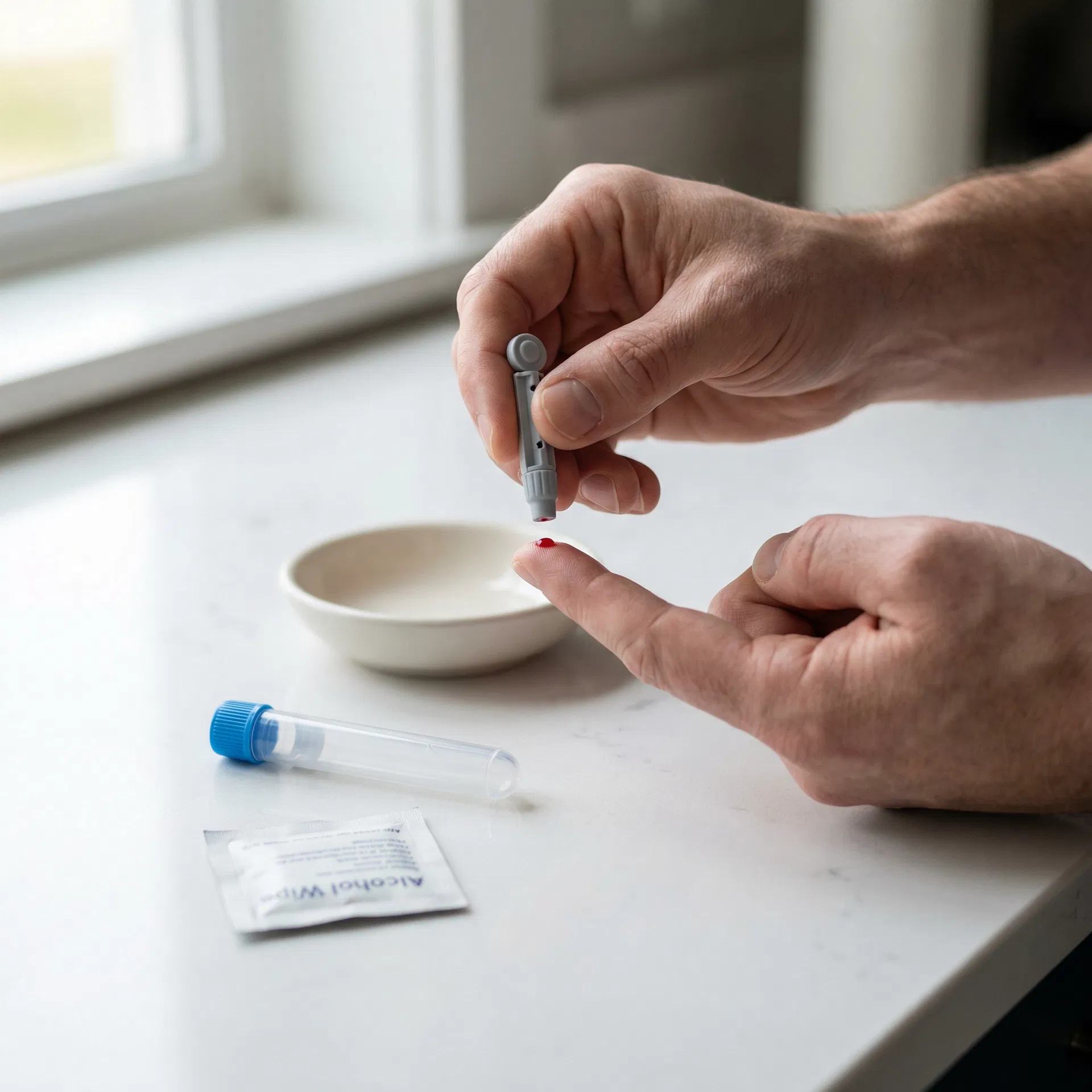
Collect your sample
Follow the simple instructions in your kit — whether it's a finger-prick at home or a venous draw at a partner clinic.
 04
04
Insights delivered
Clear, easy-to-understand results sent to you online with actionable health guidance.
Frequently asked questions
This test measures Platelet Count, MPV (Mean Platelet Volume), hs-CRP (High-Sensitivity C-Reactive Protein), Iron (Serum), TIBC (Total Iron Binding Capacity). Check the full biomarker list on this page for details.
Check Special Instructions on this page. General rule: fast 8-12 hours if cholesterol/glucose/insulin included. Most hormone, vitamin, and antibody tests do not require fasting. Morning collection (7-10am) preferred.
Follow kit instructions. Finger-prick: warm hands, use lancet as directed, fill tube to marked line. Venous: attend phlebotomy with lab form. Post same day, avoid Fridays/bank holidays.
Within the timeframe shown on this page. Notification sent when ready to view online.
You may also be interested in

Health and Lifestyle Blood Test Kit
A 19-biomarker venous blood panel covering cholesterol status, inflammation, iron status and other key health markers — ideal for investigating relevant symptoms or monitoring health.
Results estimated in 3 working days
19 biomarkers

Essential Blood Test
A 38-biomarker venous blood panel covering cholesterol status, clotting status, diabetes and other key health markers — ideal for a complete general health check across multiple body systems.
Results estimated in 3 working days
38 biomarkers

Core Health Blood Test Kit
A 7-biomarker venous blood panel covering cholesterol status and diabetes — ideal for investigating relevant symptoms or monitoring health.
Results estimated in 3 working days
7 biomarkers