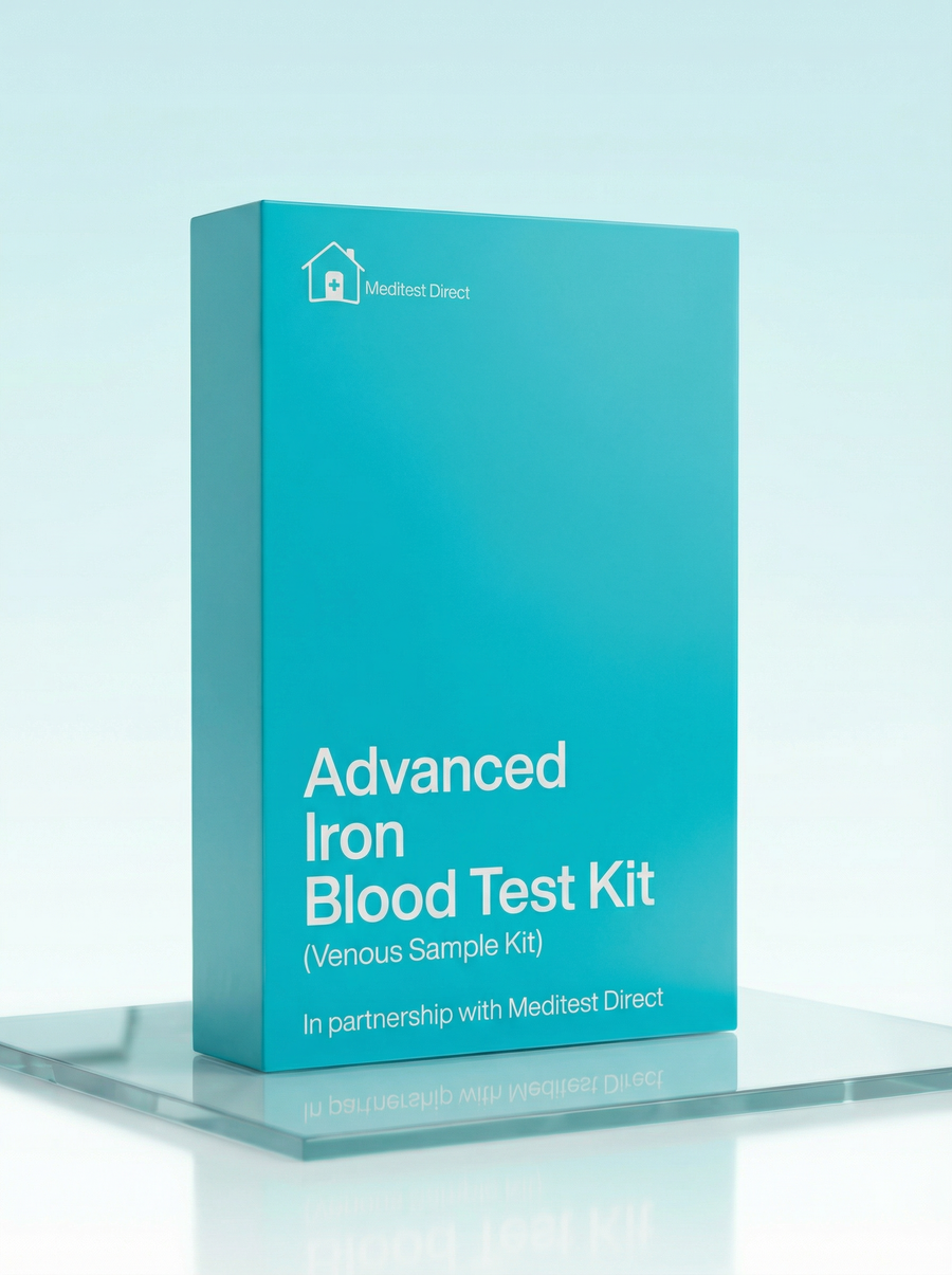
Advanced Iron Blood Test Kit
£119 ✓ In Stock
Your sample goes to a UKAS accredited laboratory meeting ISO 15189 standards.
After you receive your order confirmation email, please reply with your date of birth.
How it works
Your testing journey
From order to results in four simple steps. Full transparency on where each step happens and what it costs.

Receive your kit by post
Dispatched same working day if ordered before 3pm. Royal Mail Tracked delivery, typically 1–3 working days. 90% of kits arrive within 24 hours.

Visit a partner clinic
Book a phlebotomy appointment at one of our 365+ UK partner clinics. Take your kit with you — the phlebotomist will collect your sample using the materials provided.
Phlebotomy fee applies (paid at clinic)
Venous blood draw at a clinic
A trained phlebotomist takes a small blood sample from a vein in your arm using the vacutainers provided in your kit. The appointment takes around 10 minutes.

Return by prepaid envelope
Seal your sample in the biohazard bag provided and drop it in any Royal Mail postbox using the prepaid Tracked 24 envelope. Post Monday–Thursday for best results.
Venous Blood Collection Kit
This kit is sent to you and taken to your chosen clinic. The phlebotomist will collect your sample using the materials provided.
- 1Vacutainer blood collection tubes
- 2Needle and butterfly needle
- 3Tourniquet
- 4Alcohol swab
- 5Cotton wool and gauze
- 6Adhesive plaster
- 7Biohazard specimen bag
- 8Prepaid return envelope (Royal Mail Tracked 24)
- 9Laboratory request form
- 10Instructions for the phlebotomist
Total protein measures all the protein in your blood—mainly albumin (made by your liver) and globulins (including antibodies from your immune system). It gives a general overview of nutritional status and can hint at liver, kidney, or immune issues. Low total protein might suggest malnutrition, malabsorption, liver disease, or protein-losing conditions—all of which can coexist with iron deficiency. High total protein can occur with chronic inflammation, infection, or certain blood disorders. Protein levels provide context for interpreting other results. Results outside the normal range may need a follow-up with your GP.
Albumin is the most abundant protein in your blood, manufactured by your liver. It maintains fluid balance (keeping blood in your vessels rather than leaking into tissues), transports hormones, vitamins, and medications, and serves as a marker of nutritional status. Low albumin can indicate liver disease, kidney disease (where it leaks into urine), malnutrition, or chronic inflammation. Chronic inflammation reduces albumin production while increasing ferritin—so low albumin alongside elevated ferritin and hs-CRP suggests inflammation rather than iron overload. Albumin helps put ferritin results in context. Results outside the normal range may need a follow-up with your GP.
Globulins are a diverse family of blood proteins—some transport hormones and metals, while others (immunoglobulins) are the antibodies your immune system makes. Globulin is usually calculated by subtracting albumin from total protein. High globulins suggest your immune system is active—chronic infections, inflammation, autoimmune conditions, or liver disease. The albumin-to-globulin ratio helps interpret results: low albumin with high globulin often indicates chronic inflammation or liver disease. This is relevant to iron studies because chronic inflammation elevates ferritin independently of iron stores. Results outside the normal range may need a follow-up with your GP.
TSH (Thyroid Stimulating Hormone) is made by your pituitary gland and controls thyroid hormone production. It's included in this iron panel because thyroid function and iron metabolism are connected. Hypothyroidism (high TSH) can cause anaemia—the thyroid hormones are needed for normal red blood cell production, and hypothyroidism also reduces iron absorption. Hyperthyroidism can cause high ferritin. Additionally, iron deficiency can impair thyroid function by affecting the enzyme that makes thyroid hormones. Checking thyroid function helps explain unexplained anaemia or unusual iron results. Results outside the normal range may need a follow-up with your GP.
Free T3 is the most active thyroid hormone—the one that actually does the work in your cells, controlling metabolism and energy production. Most T3 is made by converting T4 in your tissues. Iron is required for the enzyme (thyroid peroxidase) that helps make thyroid hormones, so severe iron deficiency can impair thyroid function. Conversely, thyroid dysfunction affects red blood cell production and iron metabolism. Low Free T3 with normal T4 can indicate conversion problems or non-thyroidal illness. Including Free T3 gives the complete thyroid picture. Results outside the normal range may need a follow-up with your GP.
Free T4 is the unbound, active form of thyroxine—the main hormone your thyroid produces. It's a storage and transport hormone that gets converted to the more active T3 in tissues. Measuring Free T4 alongside TSH confirms thyroid dysfunction. Low Free T4 with high TSH confirms hypothyroidism, which can cause anaemia and affect iron absorption. High Free T4 with low TSH confirms hyperthyroidism, which can elevate ferritin. Thyroid function is checked in comprehensive iron panels because the two systems are interrelated—addressing thyroid problems can improve anaemia that doesn't respond to iron alone. Results outside the normal range may need a follow-up with your GP.
Serum iron measures the amount of iron circulating in your blood right now, bound to the transport protein transferrin. Your body needs iron to make haemoglobin (the oxygen-carrying protein in red blood cells), myoglobin (which stores oxygen in muscles), and various enzymes involved in energy production. Serum iron fluctuates considerably throughout the day—it's typically highest in the morning and can drop by 30-50% by evening. It also rises dramatically after iron-rich meals or taking iron supplements. This variability means serum iron is best interpreted alongside ferritin, TIBC, and transferrin saturation rather than on its own. Low serum iron occurs in iron deficiency but also in chronic disease and inflammation. High serum iron can indicate iron overload or recent iron ingestion. Results outside the normal range may need a follow-up with your GP.
Ferritin is the protein that stores iron inside your cells, and blood ferritin reflects those stores—it's the best single marker of your body's total iron reserves. Think of it as your iron savings account. Low ferritin is often the first sign of depleting iron stores, appearing before you become anaemic and before serum iron drops. It's common in women with heavy periods, vegetarians, endurance athletes, and people with absorption problems. The tricky part is that ferritin is also an acute phase reactant—it rises with inflammation, infection, liver disease, and chronic illness. This means a 'normal' ferritin doesn't rule out iron deficiency if you have inflammation (check your hs-CRP). Very high ferritin can indicate iron overload conditions like haemochromatosis, or it can reflect significant inflammation. Optimal ferritin for wellbeing is typically higher than the lower reference range. Results outside the normal range may need a follow-up with your GP.
TIBC measures the total capacity of your blood to bind and transport iron—essentially, it reflects how much transferrin (the iron transport protein) is available. Think of it as counting the total 'seats' available on the iron transport system. When iron stores are low, your liver makes more transferrin to capture every bit of iron it can, so TIBC goes up—this is a compensatory response to iron deficiency. When iron stores are high, there's less need for transport capacity, so TIBC drops. High TIBC with low serum iron and low ferritin is the classic pattern of iron deficiency anaemia. Low TIBC can occur with iron overload, chronic disease, malnutrition, and liver problems. TIBC is more stable than serum iron throughout the day, making it a more reliable marker. Results outside the normal range may need a follow-up with your GP.
UIBC measures the reserve capacity of transferrin that isn't currently carrying iron—the 'empty seats' on the iron transport system. Together with serum iron, UIBC can be used to calculate TIBC (serum iron + UIBC = TIBC). In iron deficiency, UIBC is high because there's lots of unused capacity—your transferrin has empty binding sites waiting for iron that isn't there. In iron overload, UIBC is low because most binding sites are already occupied. Some labs measure TIBC directly, others calculate it from UIBC and serum iron—the information is equivalent. UIBC helps complete the picture of your iron transport system and can be particularly useful for identifying iron deficiency in the early stages. Results outside the normal range may need a follow-up with your GP.
Transferrin saturation tells you what percentage of your iron transport capacity is currently being used—calculated by dividing serum iron by TIBC. It's one of the most useful markers for both iron deficiency and iron overload. Normal saturation is typically 20-45%. Low saturation (below 20%) means lots of empty 'seats' on your transferrin—this strongly suggests iron deficiency, especially when combined with low ferritin. High saturation (above 45%) means most binding sites are full—this can indicate iron overload. In haemochromatosis (genetic iron overload), transferrin saturation is often one of the first markers to become abnormal, sometimes rising before ferritin does. Very high saturation (above 60%) is concerning for significant iron overload. Because it's a ratio, transferrin saturation accounts for individual variation in transferrin production. Results outside the normal range may need a follow-up with your GP.
Bilirubin is the yellow pigment produced when red blood cells break down. Your liver processes bilirubin and excretes it in bile. Elevated bilirubin causes jaundice—yellowing of the skin and eyes. In the context of iron studies, bilirubin can help identify haemolytic anaemia (where red cells are being destroyed faster than normal), which raises bilirubin while causing anaemia. High bilirubin can also indicate liver disease or bile duct obstruction. A common cause of mildly elevated bilirubin is Gilbert's syndrome—a benign genetic condition affecting about 5% of people where the liver is slower at processing bilirubin. Results outside the normal range may need a follow-up with your GP.
ALP is an enzyme found mainly in your liver and bones, with smaller amounts in kidneys and gut. In the liver, it's concentrated in cells lining the bile ducts. Elevated ALP can indicate bile duct problems (cholestasis), liver disease, or bone conditions. It's relevant to iron studies because iron overload conditions like haemochromatosis can cause liver damage and raised ALP. ALP is naturally higher in children and teenagers (growing bones) and during pregnancy. When ALP is elevated, checking GGT helps determine whether the source is liver (GGT also elevated) or bone (GGT normal). Results outside the normal range may need a follow-up with your GP.
ALT is an enzyme that lives mainly inside your liver cells. When those cells are damaged or inflamed, ALT leaks into your bloodstream—so elevated levels signal liver cell injury. This is important in iron studies because iron overload (haemochromatosis) causes iron to deposit in the liver, leading to damage and raised ALT over time. Fatty liver disease, alcohol, viral hepatitis, and certain medications also raise ALT. In someone with high ferritin and high transferrin saturation, elevated ALT adds evidence for iron overload affecting the liver and increases the urgency for investigation. Results outside the normal range may need a follow-up with your GP.
GGT is an enzyme concentrated in your liver and bile ducts. It's particularly sensitive to alcohol—even moderate regular drinking can push it up—making it useful for spotting alcohol-related liver effects. GGT also rises with bile duct problems, fatty liver, and certain medications. When ALP is elevated, GGT helps identify whether the source is liver (GGT also elevated) or bone (GGT normal, since bones don't contain GGT). In the context of iron overload, elevated GGT alongside raised ALT supports the presence of liver involvement. An isolated raised GGT often prompts a look at alcohol intake. Results outside the normal range may need a follow-up with your GP.
Urea is a waste product formed when your liver breaks down protein. It travels through your blood to your kidneys, which filter it out into urine. Kidney function is relevant to iron studies because the kidneys produce erythropoietin (EPO), the hormone that stimulates red blood cell production. Chronic kidney disease leads to reduced EPO, causing anaemia that doesn't respond to iron supplementation alone. High urea with normal creatinine often suggests dehydration or high protein intake. High urea with high creatinine and low eGFR indicates kidney dysfunction. Results outside the normal range may need a follow-up with your GP.
Creatinine is a waste product from the normal wear and tear of your muscles. Your body produces it at a fairly steady rate, and healthy kidneys filter it out into urine. Because production is consistent and clearance depends on kidney function, creatinine is a reliable marker for how well your kidneys are working. Higher levels suggest reduced kidney filtering efficiency. Creatinine is affected by muscle mass—muscular people naturally run higher. In the context of iron studies, creatinine helps identify kidney disease as a potential cause of anaemia. Results outside the normal range may need a follow-up with your GP.
eGFR (estimated Glomerular Filtration Rate) calculates how much blood your kidneys filter per minute—essentially measuring their efficiency. It's worked out from your creatinine along with age, sex, and ethnicity. A higher number is better: above 90 is generally normal, while lower numbers indicate declining kidney function. The beauty of eGFR is that it can detect kidney problems earlier than creatinine alone. Kidney disease causes anaemia through reduced EPO production, so low eGFR in someone with anaemia might explain why the anaemia persists despite adequate iron. Results outside the normal range may need a follow-up with your GP.
Sodium is an essential electrolyte that regulates fluid balance, blood pressure, and nerve/muscle function. Your kidneys carefully control sodium levels. Low sodium (hyponatraemia) can occur with excessive water intake, certain medications, heart failure, liver disease, and kidney problems—symptoms include confusion, fatigue, and headaches. High sodium (hypernatraemia) usually indicates dehydration or excessive salt intake. While not directly related to iron metabolism, sodium helps assess overall kidney function and hydration status, which can affect how other results are interpreted. Results outside the normal range may need a follow-up with your GP.
Folate (vitamin B9) is essential for DNA synthesis and red blood cell production—it works alongside iron and B12 to produce healthy red cells. Folate deficiency causes macrocytic anaemia (large cells, high MCV) in contrast to iron deficiency's microcytic anaemia (small cells, low MCV). Sometimes people have combined deficiencies—the MCV might be misleadingly normal because the opposing effects cancel out. Good dietary sources include leafy greens, legumes, and fortified cereals. Low folate can be caused by poor diet, excessive alcohol, certain medications, or malabsorption conditions like coeliac disease. Checking folate alongside iron gives a complete picture of nutrients needed for blood production. Results outside the normal range may need a follow-up with your GP.
Active B12 (holotranscobalamin) measures the form of B12 your body can actually use. Like folate, B12 is essential for red blood cell production and DNA synthesis. B12 deficiency causes macrocytic anaemia with large, immature red cells. It can coexist with iron deficiency—someone might have combined deficiencies from poor absorption (coeliac disease, gastric surgery) or restricted diets. Vegans and vegetarians are at higher risk for B12 deficiency since it comes mainly from animal foods. B12 deficiency also causes neurological symptoms—pins and needles, memory problems, mood changes—that iron deficiency doesn't. Checking B12 alongside iron ensures you're not missing another cause of anaemia. Results outside the normal range may need a follow-up with your GP.
Haemoglobin is the iron-containing protein inside red blood cells that carries oxygen from your lungs to every tissue in your body. It's what makes blood red. Low haemoglobin—anaemia—is the end result of prolonged iron deficiency: once your iron stores are depleted and there's not enough iron to make new haemoglobin, your haemoglobin level drops. Symptoms include fatigue, weakness, shortness of breath, dizziness, and pale skin. By the time haemoglobin falls, iron deficiency is typically quite advanced. Other causes of low haemoglobin include B12 or folate deficiency, chronic disease, kidney disease, and blood loss. High haemoglobin can occur with dehydration, living at altitude, lung disease, or conditions that overproduce red blood cells. Results outside the normal range may need a follow-up with your GP.
Haematocrit measures what percentage of your blood volume is made up of red blood cells. If your haematocrit is 42%, that means 42% of your blood is red cells and the rest is plasma plus other cells. It tracks closely with haemoglobin—low haematocrit usually indicates anaemia, high haematocrit can indicate dehydration or overproduction of red cells. In iron deficiency anaemia, haematocrit falls as the body produces fewer and smaller red cells. Haematocrit is useful for assessing blood viscosity and oxygen-carrying capacity. Athletes may have higher haematocrit from training adaptations. Very high haematocrit increases blood viscosity and clotting risk. Results outside the normal range may need a follow-up with your GP.
Red cell count measures the actual number of red blood cells in a volume of your blood. Red cells are produced in your bone marrow and live for about 120 days before being recycled. In iron deficiency, the bone marrow can still produce red cells, but they're smaller and contain less haemoglobin—so the red cell count may be relatively preserved while MCV and MCH fall. In other types of anaemia (B12/folate deficiency), red cells are large but fewer in number. Low red cell count can indicate anaemia from various causes, while high red cell count can occur with dehydration, lung disease, or bone marrow conditions. Results outside the normal range may need a follow-up with your GP.
MCV measures the average size of your red blood cells and is one of the most useful markers for identifying the type of anaemia. In iron deficiency, cells are smaller than normal (low MCV, 'microcytic')—there isn't enough iron to fill them properly, so the bone marrow produces smaller cells. This is the hallmark of iron deficiency anaemia, also seen in thalassaemia. In B12 or folate deficiency, cells are larger than normal (high MCV, 'macrocytic')—DNA synthesis is impaired so cells grow larger before dividing. Normal-sized cells with low haemoglobin ('normocytic' anaemia) can occur with chronic disease, kidney problems, or recent blood loss. MCV helps guide what additional tests might be needed. Results outside the normal range may need a follow-up with your GP.
MCH tells you the average amount of haemoglobin inside each red blood cell—how 'full' of haemoglobin your red cells are. In iron deficiency, cells contain less haemoglobin than normal (low MCH) because there isn't enough iron to make adequate haemoglobin—producing pale, 'hypochromic' cells. Low MCH typically accompanies low MCV in iron deficiency. High MCH suggests larger-than-normal cells packed with haemoglobin, typically seen with B12 or folate deficiency. MCH helps quantify the severity of iron deficiency and track response to treatment—as iron stores replenish, MCH should gradually normalise. Results outside the normal range may need a follow-up with your GP.
MCHC measures the concentration of haemoglobin within your red blood cells—how densely packed with haemoglobin they are, accounting for cell size. While MCH looks at the total amount of haemoglobin per cell, MCHC tells you how concentrated it is. Low MCHC produces 'hypochromic' (pale) cells—a classic finding in iron deficiency where cells can't be filled properly with haemoglobin. MCHC is one of the last red cell indices to become abnormal in iron deficiency and can help confirm the diagnosis. Very high MCHC is rare but can occur in conditions where red cells are abnormally shaped. Results outside the normal range may need a follow-up with your GP.
RDW measures how much variation there is in the size of your red blood cells. Normally, red cells are fairly uniform in size, so a low RDW is good. A high RDW means you have a mix of different-sized cells—some big, some small—which can happen when your bone marrow is producing abnormal cells or when you have mixed causes of anaemia. In iron deficiency, RDW often rises early—sometimes before MCV falls—as some normal-sized cells remain while new smaller cells are being produced. This makes RDW useful for detecting early iron deficiency. High RDW with low MCV strongly suggests iron deficiency. High RDW can also occur with B12/folate deficiency, mixed deficiencies, or after blood transfusion. Results outside the normal range may need a follow-up with your GP.
White blood cells are your immune system's soldiers, defending you against infections, viruses, and other threats. The total white cell count gives an overall picture of immune activity. Elevated counts often signal that your immune system is fighting something—infection, inflammation, or stress. This is relevant for interpreting iron results because infections and inflammation affect iron markers (especially ferritin). A very high white count can indicate serious infection or occasionally blood disorders. Low white count can occur with viral infections, certain medications, autoimmune conditions, or bone marrow problems. Results outside the normal range may need a follow-up with your GP.
Neutrophils are the most abundant type of white blood cell and your first responders to bacterial infections. They're like the infantry of your immune system—quick to arrive at infection sites where they engulf and destroy bacteria. High neutrophils often indicate bacterial infection or acute inflammation. Low neutrophils (neutropenia) can occur with viral infections, certain medications (especially chemotherapy), or bone marrow problems—it increases your risk of bacterial infections. Elevated neutrophils alongside elevated hs-CRP suggest active inflammation, which is important context for interpreting ferritin results. Results outside the normal range may need a follow-up with your GP.
Lymphocytes handle the more sophisticated parts of immunity—they include T-cells (which kill infected cells and coordinate immune responses), B-cells (which make antibodies), and natural killer cells. High lymphocytes often occur with viral infections—your body's fighting the virus—and some autoimmune conditions. Low lymphocytes can occur with HIV, after chemotherapy, or with severe infections. The balance between neutrophils and lymphocytes can help distinguish viral from bacterial infections. Chronic lymphocyte elevation might indicate ongoing immune stimulation. Results outside the normal range may need a follow-up with your GP.
Monocytes are white blood cells that travel through your bloodstream and then move into tissues where they become macrophages—large cells that engulf and digest bacteria, dead cells, and debris. They're the cleanup crew after an infection. High monocytes can occur with chronic infections (like tuberculosis), autoimmune conditions, and recovery from acute infections. Monocytes also play a role in iron regulation—macrophages recycle iron from old red blood cells and can sequester iron during inflammation, contributing to the anaemia of chronic disease. Results outside the normal range may need a follow-up with your GP.
Eosinophils are white blood cells particularly involved in fighting parasites and in allergic reactions. High eosinophils (eosinophilia) commonly occur with allergies, asthma, eczema, hay fever, and parasitic infections. They can also be elevated with certain autoimmune conditions and some blood disorders. If you have seasonal allergies, don't be surprised to see elevated eosinophils during pollen season. Parasitic infections can cause iron deficiency through blood loss or malabsorption, so elevated eosinophils alongside iron deficiency might prompt investigation for parasites. Results outside the normal range may need a follow-up with your GP.
Basophils are the rarest type of white blood cell, making up less than 1% of your total white cells. They play a role in inflammatory and allergic responses, releasing histamine and other chemicals that contribute to allergic symptoms. Elevated basophils can occur with allergic reactions, hypothyroidism, and some blood disorders. Because they're so few in number, small changes can look significant on a percentage basis. Isolated mild basophil elevation is rarely concerning. Results outside the normal range may need a follow-up with your GP.
hs-CRP is perhaps the most important supporting marker in this panel for interpreting iron results correctly. CRP is produced by your liver in response to inflammation anywhere in your body. The high-sensitivity version can detect low-grade chronic inflammation. Ferritin is an acute phase reactant—it rises with inflammation regardless of iron stores. This means someone with iron deficiency AND inflammation might have a 'normal' ferritin that's actually masking depleted iron stores. If your hs-CRP is elevated, your ferritin might be falsely reassuring. Conversely, if hs-CRP is normal and ferritin is high, you're more likely dealing with genuine iron overload rather than inflammation. hs-CRP helps interpret ferritin accurately. Results outside the normal range may need a follow-up with your GP.
Platelets are tiny cell fragments that clump together to form clots and stop bleeding. Interestingly, platelet count can be elevated in iron deficiency—this reactive thrombocytosis is thought to be a compensatory response, though the mechanism isn't entirely clear. High platelets can also occur with inflammation, infection, and after splenectomy. Low platelets (thrombocytopenia) can mean bruising easily and bleeding that's hard to stop—causes include viral infections, certain medications, autoimmune conditions, and bone marrow problems. Very low platelet counts need urgent investigation. Results outside the normal range may need a follow-up with your GP.
MPV measures the average size of your platelets. Larger platelets are typically younger and more active—they've just been released from the bone marrow and are more effective at clotting. High MPV with low platelet count suggests your bone marrow is working hard to produce new platelets to replace those being destroyed—seen in conditions like immune thrombocytopenia. Low MPV with low platelet count suggests the bone marrow itself isn't producing enough. In iron deficiency with reactive thrombocytosis, MPV is often normal or slightly decreased. MPV helps interpret platelet count abnormalities. Results outside the normal range may need a follow-up with your GP.
This test is for screening and information only — it is not a medical diagnosis or professional advice. Please have your results reviewed by a qualified doctor or healthcare provider who can explain what they mean for your personal health situation. If your results show anything outside the normal range, or if you're worried about your health, see your doctor as soon as you can. Don't change any medications or treatments based on these results alone — always talk to your healthcare provider first.
NO CLINICS, NO QUEUES, NO HASSLE
Four steps to clarity
 01
01
Pick your panel
Browse over 200 clinically designed test kits and choose the one that fits your goals.
 02
02
Kit to your door
Everything you need arrives in discreet packaging with step-by-step instructions inside.
 03
03
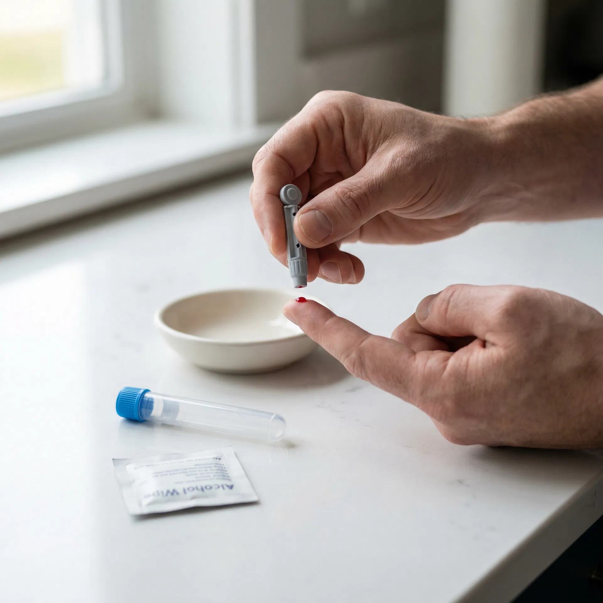
Collect your sample
Follow the simple instructions in your kit — whether it's a finger-prick at home or a venous draw at a partner clinic.
 04
04
Insights delivered
Clear, easy-to-understand results sent to you online with actionable health guidance.
Frequently asked questions
This test measures Iron, Ferritin, TIBC (Total Iron Binding Capacity), UIBC (Unsaturated Iron Binding Capacity), Transferrin Saturation. Check the full biomarker list on this page for detailed descriptions.
Check the Special Instructions on this page. General rule: fast 8-12 hours if cholesterol/glucose/insulin included. Most hormone, vitamin, and antibody tests do not require fasting. Morning collection (7-10am) is preferred.
Follow the instructions in your kit. For finger-prick: warm hands, use lancet as directed, fill tube to marked line. For venous: attend a phlebotomy clinic with your lab form. Post same day, avoid Fridays/bank holidays.
Results are typically available within the timeframe shown on this page. You will receive a notification when ready to view online.
You may also be interested in

NAD+ Blood Test
A 2-biomarker venous blood panel covering cellular energy & longevity — ideal for optimising cellular energy production and longevity.
Results estimated in 7–10 working days
2 biomarkers

Vitamin B2 (riboflavin) Blood Test
A single-biomarker venous blood test covering vitamins — ideal for investigating B vitamin deficiency or nervous system symptoms.
Results estimated in 9 working days
1 biomarkers

Vitamin B9 (folic acid) Blood Test Kit
A single-biomarker venous blood test covering vitamins — ideal for investigating folate deficiency, fatigue, or pre-conception health.
Results estimated in 2 working days
1 biomarkers

Driver's Tiredness Blood Test
A 6-biomarker venous blood panel covering inflammation, iron status, thyroid hormones and vitamins — ideal for investigating ongoing tiredness, low energy, or suspected medical causes.
Results estimated in 5 working days
6 biomarkers